Credits and licences
Every note, image and 3D model on Anatomy Interface comes from an openly licensed source. Each one shows its own credit where it appears: its title, its author, a link to the exact source page and a link to its licence. This page lists every source the same way, grouped by type.
Reusing what you find here. The notes, images and models keep their original licences (CC BY-SA, CC BY, CC0 or public domain) and can be reused under those terms, with the credit shown for each one. The 3D models we adapted are shared under CC BY-SA 4.0, as the Z-Anatomy licence requires.
The site's own code, design and tools are not openly licensed.
To report a wrong or missing credit, use the Report a mistake link at the bottom of this page.
3D models
| Title (linked to the source) | Author | Licence | Used for |
|---|---|---|---|
| Z-Anatomy - The libre 3D atlas of anatomy (the wording the Z-Anatomy Readme asks for: "Z-Anatomy - The libre 3D atlas of anatomy - CC-BY-SA 4.0") | Gauthier Kervyn (design, 3D, anatomy) and Marcin Zielinski (Python script), as the Z-Anatomy Readme names them | CC BY-SA 4.0 | Every 3D model except the skin surface, the renal pelvis and the inner ear; all structure names. Adapted: split into layers and pages, compressed, some parts removed or replaced. |
| BodyParts3D ("BodyParts3D - The Database Center for Life Science - CC-BY-SA 2.1 Japan"), the source Z-Anatomy's meshes are derived from | Database Center for Life Science (DBCLS), Japan; original model by Kousaku Okubo (as the Z-Anatomy Readme names him) | CC BY-SA 2.1 Japan, as Z-Anatomy states it (DBCLS's own licence page now says CC BY 4.0) | Underlying meshes |
| 3D Reference Organ for Renal Pelvis, Male, Left v1.0, Human Reference Atlas (HuBMAP), NIH 3D 3DPX-021623; from Visible Human Male data, courtesy of the U.S. National Library of Medicine | Kristen Browne, Heidi Schlehlein | CC BY 4.0 | Left renal pelvis on the urinary page. Moved and scaled to fit; position approximate. |
| 3D Reference Organ for Renal Pelvis, Male, Right v1.0, Human Reference Atlas (HuBMAP), NIH 3D 3DPX-021624; from Visible Human Male data, courtesy of the U.S. National Library of Medicine | Kristen Browne, Heidi Schlehlein | CC BY 4.0 | Right renal pelvis on the urinary page. Moved and scaled to fit; position approximate. |
IE-Map: a novel in-vivo atlas and template of the human inner ear (data v1.0.0, files mesh_david_cochlea.vtk and segT2_th1.07_Otsu.vtk; paper: Sci Rep 11, 3293, 2021) | Seyed-Ahmad Ahmadi, Theresa Marie Raiser, Ria Maxine Rühl, Virginia Flanagin, Peter zu Eulenburg; German Center for Vertigo and Balance Disorders | CC BY 4.0 | Cochlea and inner ear labyrinth on the nervous and eye and ear pages. Left ear mirrored; placed by fitting; position approximate. |
| MPFB2 / MakeHuman base mesh and targets | MakeHuman community | CC0 1.0 | The skin surface of the homepage body |
Other models inside Z-Anatomy (the known gap)
The Z-Anatomy Readme lists other models it draws on, but doesn't record which parts of the dataset come from which:
- "Cranial Nerves and Foramina" by the University of Dundee, CAHID (produced by Sophia Lappe; its page says it is based on BodyParts3D), CC BY 4.0 as stated in the Z-Anatomy Readme (the model's own page states no licence today). Some cranial nerves may come from it, so every page that shows a cranial nerve credits it: the homepage and the nervous system, eye and ear, head and neck, thorax and abdomen pages.
- "Brainder" and "White matter" (University of Washington): licence not stated. Some brain parts may come from them; BodyParts3D, which is openly licensed, also has the cerebral gyri and white matter.
- "Anatomy of the Inner Ear", University of Dundee, CC BY-NC-SA 4.0 (non-commercial), and "Kidney" by Lissie Cowley, CC BY-NC 4.0 (non-commercial). To be safe, the parts that could come from these were replaced or removed: the renal pelvis (replaced with the Human Reference Atlas), the cochlea and vestibule (replaced with IE-Map), and the malleus, incus, stapes and eardrum (removed). The kidneys themselves match BodyParts3D and stay.
Notes on the 3D parts
Short excerpts, copied word for word. Each note on the site shows the same credit as its line below.
- Wikipedia, through the Z-Anatomy definitions: "Article" by Wikipedia contributors, via the Z-Anatomy definitions, CC BY-SA 3.0 (as the Z-Anatomy Readme states).
- Wikipedia article leads fetched directly (8 to 12 Nov 2026): "Article" by Wikipedia contributors, CC BY-SA 4.0. Matching a part to its article used Wikidata (CC0); only Wikidata and FMA ID numbers are kept, no FMA text.
The authors of each Wikipedia article are listed in its history (the "View history" tab of each article).
Wikipedia articles quoted under CC BY-SA 3.0, via the Z-Anatomy definitions (932)
- "Abdominal aorta" by Wikipedia contributors, via the Z-Anatomy definitions, CC BY-SA 3.0
- "Abdominal external oblique muscle" by Wikipedia contributors, via the Z-Anatomy definitions, CC BY-SA 3.0
- "Abdominal fascia" by Wikipedia contributors, via the Z-Anatomy definitions, CC BY-SA 3.0
- "Abdominal internal oblique muscle" by Wikipedia contributors, via the Z-Anatomy definitions, CC BY-SA 3.0
- "Abducens nerve" by Wikipedia contributors, via the Z-Anatomy definitions, CC BY-SA 3.0
- "Abducens nucleus" by Wikipedia contributors, via the Z-Anatomy definitions, CC BY-SA 3.0
- "Abductor digiti minimi muscle of foot" by Wikipedia contributors, via the Z-Anatomy definitions, CC BY-SA 3.0
- "Abductor digiti minimi muscle of hand" by Wikipedia contributors, via the Z-Anatomy definitions, CC BY-SA 3.0
- "Abductor hallucis muscle" by Wikipedia contributors, via the Z-Anatomy definitions, CC BY-SA 3.0
- "Abductor pollicis brevis muscle" by Wikipedia contributors, via the Z-Anatomy definitions, CC BY-SA 3.0
- "Abductor pollicis longus muscle" by Wikipedia contributors, via the Z-Anatomy definitions, CC BY-SA 3.0
- "Accessory nerve" by Wikipedia contributors, via the Z-Anatomy definitions, CC BY-SA 3.0
- "Acetabular labrum" by Wikipedia contributors, via the Z-Anatomy definitions, CC BY-SA 3.0
- "Achilles tendon" by Wikipedia contributors, via the Z-Anatomy definitions, CC BY-SA 3.0
- "Acromioclavicular joint" by Wikipedia contributors, via the Z-Anatomy definitions, CC BY-SA 3.0
- "Acromioclavicular ligament" by Wikipedia contributors, via the Z-Anatomy definitions, CC BY-SA 3.0
- "Adductor brevis muscle" by Wikipedia contributors, via the Z-Anatomy definitions, CC BY-SA 3.0
- "Adductor hallucis muscle" by Wikipedia contributors, via the Z-Anatomy definitions, CC BY-SA 3.0
- "Adductor longus muscle" by Wikipedia contributors, via the Z-Anatomy definitions, CC BY-SA 3.0
- "Adductor magnus muscle" by Wikipedia contributors, via the Z-Anatomy definitions, CC BY-SA 3.0
- "Adductor minimus muscle" by Wikipedia contributors, via the Z-Anatomy definitions, CC BY-SA 3.0
- "Adductor pollicis muscle" by Wikipedia contributors, via the Z-Anatomy definitions, CC BY-SA 3.0
- "Amygdala" by Wikipedia contributors, via the Z-Anatomy definitions, CC BY-SA 3.0
- "Anconeus muscle" by Wikipedia contributors, via the Z-Anatomy definitions, CC BY-SA 3.0
- "Angular artery" by Wikipedia contributors, via the Z-Anatomy definitions, CC BY-SA 3.0
- "Angular vein" by Wikipedia contributors, via the Z-Anatomy definitions, CC BY-SA 3.0
- "Annular ligament of radius" by Wikipedia contributors, via the Z-Anatomy definitions, CC BY-SA 3.0
- "Anserine bursa" by Wikipedia contributors, via the Z-Anatomy definitions, CC BY-SA 3.0
- "Antebrachial fascia" by Wikipedia contributors, via the Z-Anatomy definitions, CC BY-SA 3.0
- "Anterior branch of obturator nerve" by Wikipedia contributors, via the Z-Anatomy definitions, CC BY-SA 3.0
- "Anterior cerebral artery" by Wikipedia contributors, via the Z-Anatomy definitions, CC BY-SA 3.0
- "Anterior chamber of eyeball" by Wikipedia contributors, via the Z-Anatomy definitions, CC BY-SA 3.0
- "Anterior commissure" by Wikipedia contributors, via the Z-Anatomy definitions, CC BY-SA 3.0
- "Anterior communicating artery" by Wikipedia contributors, via the Z-Anatomy definitions, CC BY-SA 3.0
- "Anterior corticospinal tract" by Wikipedia contributors, via the Z-Anatomy definitions, CC BY-SA 3.0
- "Anterior cruciate ligament" by Wikipedia contributors, via the Z-Anatomy definitions, CC BY-SA 3.0
- "Anterior ethmoidal artery" by Wikipedia contributors, via the Z-Anatomy definitions, CC BY-SA 3.0
- "Anterior grey column" by Wikipedia contributors, via the Z-Anatomy definitions, CC BY-SA 3.0
- "Anterior inferior cerebellar artery" by Wikipedia contributors, via the Z-Anatomy definitions, CC BY-SA 3.0
- "Anterior intermuscular septum of leg" by Wikipedia contributors, via the Z-Anatomy definitions, CC BY-SA 3.0
- "Anterior interosseous nerve" by Wikipedia contributors, via the Z-Anatomy definitions, CC BY-SA 3.0
- "Anterior jugular vein" by Wikipedia contributors, via the Z-Anatomy definitions, CC BY-SA 3.0
- "Anterior ligament of the head of the fibula" by Wikipedia contributors, via the Z-Anatomy definitions, CC BY-SA 3.0
- "Anterior longitudinal ligament" by Wikipedia contributors, via the Z-Anatomy definitions, CC BY-SA 3.0
- "Anterior segment of eyeball" by Wikipedia contributors, via the Z-Anatomy definitions, CC BY-SA 3.0
- "Anterior spinal artery" by Wikipedia contributors, via the Z-Anatomy definitions, CC BY-SA 3.0
- "Anterior sternoclavicular ligament" by Wikipedia contributors, via the Z-Anatomy definitions, CC BY-SA 3.0
- "Anterior talocalcaneal ligament" by Wikipedia contributors, via the Z-Anatomy definitions, CC BY-SA 3.0
- "Anterior tibial artery" by Wikipedia contributors, via the Z-Anatomy definitions, CC BY-SA 3.0
- "Anterior tibial vein" by Wikipedia contributors, via the Z-Anatomy definitions, CC BY-SA 3.0
- "Anterior tibiofibular ligament" by Wikipedia contributors, via the Z-Anatomy definitions, CC BY-SA 3.0
- "Aorta" by Wikipedia contributors, via the Z-Anatomy definitions, CC BY-SA 3.0
- "Aortic arch" by Wikipedia contributors, via the Z-Anatomy definitions, CC BY-SA 3.0
- "Aortic valve" by Wikipedia contributors, via the Z-Anatomy definitions, CC BY-SA 3.0
- "Appendicular artery" by Wikipedia contributors, via the Z-Anatomy definitions, CC BY-SA 3.0
- "Appendix (anatomy)" by Wikipedia contributors, via the Z-Anatomy definitions, CC BY-SA 3.0
- "Arcuate popliteal ligament" by Wikipedia contributors, via the Z-Anatomy definitions, CC BY-SA 3.0
- "Articular disc of sternoclavicular joint" by Wikipedia contributors, via the Z-Anatomy definitions, CC BY-SA 3.0
- "Articular disk of the temporomandibular joint" by Wikipedia contributors, via the Z-Anatomy definitions, CC BY-SA 3.0
- "Arytenoid cartilage" by Wikipedia contributors, via the Z-Anatomy definitions, CC BY-SA 3.0
- "Ascending aorta" by Wikipedia contributors, via the Z-Anatomy definitions, CC BY-SA 3.0
- "Ascending pharyngeal artery" by Wikipedia contributors, via the Z-Anatomy definitions, CC BY-SA 3.0
- "Atlas (anatomy)" by Wikipedia contributors, via the Z-Anatomy definitions, CC BY-SA 3.0
- "Atrium (heart)" by Wikipedia contributors, via the Z-Anatomy definitions, CC BY-SA 3.0
- "Auricle (anatomy)" by Wikipedia contributors, via the Z-Anatomy definitions, CC BY-SA 3.0
- "Axillary artery" by Wikipedia contributors, via the Z-Anatomy definitions, CC BY-SA 3.0
- "Axillary lymph nodes" by Wikipedia contributors, via the Z-Anatomy definitions, CC BY-SA 3.0
- "Axillary nerve" by Wikipedia contributors, via the Z-Anatomy definitions, CC BY-SA 3.0
- "Axillary vein" by Wikipedia contributors, via the Z-Anatomy definitions, CC BY-SA 3.0
- "Axis (anatomy)" by Wikipedia contributors, via the Z-Anatomy definitions, CC BY-SA 3.0
- "Azygos vein" by Wikipedia contributors, via the Z-Anatomy definitions, CC BY-SA 3.0
- "Basilar artery" by Wikipedia contributors, via the Z-Anatomy definitions, CC BY-SA 3.0
- "Basilar plexus" by Wikipedia contributors, via the Z-Anatomy definitions, CC BY-SA 3.0
- "Basilic vein" by Wikipedia contributors, via the Z-Anatomy definitions, CC BY-SA 3.0
- "Biceps" by Wikipedia contributors, via the Z-Anatomy definitions, CC BY-SA 3.0
- "Biceps femoris muscle" by Wikipedia contributors, via the Z-Anatomy definitions, CC BY-SA 3.0
- "Bicipitoradial bursa" by Wikipedia contributors, via the Z-Anatomy definitions, CC BY-SA 3.0
- "Bile duct" by Wikipedia contributors, via the Z-Anatomy definitions, CC BY-SA 3.0
- "Brachial artery" by Wikipedia contributors, via the Z-Anatomy definitions, CC BY-SA 3.0
- "Brachial fascia" by Wikipedia contributors, via the Z-Anatomy definitions, CC BY-SA 3.0
- "Brachial lymph nodes" by Wikipedia contributors, via the Z-Anatomy definitions, CC BY-SA 3.0
- "Brachial plexus" by Wikipedia contributors, via the Z-Anatomy definitions, CC BY-SA 3.0
- "Brachial plexus#Roots" by Wikipedia contributors, via the Z-Anatomy definitions, CC BY-SA 3.0
- "Brachial veins" by Wikipedia contributors, via the Z-Anatomy definitions, CC BY-SA 3.0
- "Brachiocephalic vein" by Wikipedia contributors, via the Z-Anatomy definitions, CC BY-SA 3.0
- "Brachioradialis" by Wikipedia contributors, via the Z-Anatomy definitions, CC BY-SA 3.0
- "Brain" by Wikipedia contributors, via the Z-Anatomy definitions, CC BY-SA 3.0
- "Bronchus" by Wikipedia contributors, via the Z-Anatomy definitions, CC BY-SA 3.0
- "Buccal artery" by Wikipedia contributors, via the Z-Anatomy definitions, CC BY-SA 3.0
- "Buccal nerve" by Wikipedia contributors, via the Z-Anatomy definitions, CC BY-SA 3.0
- "Buccinator lymph node" by Wikipedia contributors, via the Z-Anatomy definitions, CC BY-SA 3.0
- "Buccinator muscle" by Wikipedia contributors, via the Z-Anatomy definitions, CC BY-SA 3.0
- "Calcaneocuboid ligament" by Wikipedia contributors, via the Z-Anatomy definitions, CC BY-SA 3.0
- "Calcaneofibular ligament" by Wikipedia contributors, via the Z-Anatomy definitions, CC BY-SA 3.0
- "Calcaneus" by Wikipedia contributors, via the Z-Anatomy definitions, CC BY-SA 3.0
- "Canine tooth" by Wikipedia contributors, via the Z-Anatomy definitions, CC BY-SA 3.0
- "Capitate bone" by Wikipedia contributors, via the Z-Anatomy definitions, CC BY-SA 3.0
- "Carotid triangle" by Wikipedia contributors, via the Z-Anatomy definitions, CC BY-SA 3.0
- "Cauda equina" by Wikipedia contributors, via the Z-Anatomy definitions, CC BY-SA 3.0
- "Caudate nucleus" by Wikipedia contributors, via the Z-Anatomy definitions, CC BY-SA 3.0
- "Cavernous sinus" by Wikipedia contributors, via the Z-Anatomy definitions, CC BY-SA 3.0
- "Celiac artery" by Wikipedia contributors, via the Z-Anatomy definitions, CC BY-SA 3.0
- "Central canal" by Wikipedia contributors, via the Z-Anatomy definitions, CC BY-SA 3.0
- "Central lobule" by Wikipedia contributors, via the Z-Anatomy definitions, CC BY-SA 3.0
- "Central lymph nodes" by Wikipedia contributors, via the Z-Anatomy definitions, CC BY-SA 3.0
- "Central retinal artery" by Wikipedia contributors, via the Z-Anatomy definitions, CC BY-SA 3.0
- "Cephalic vein" by Wikipedia contributors, via the Z-Anatomy definitions, CC BY-SA 3.0
- "Cerebellum" by Wikipedia contributors, via the Z-Anatomy definitions, CC BY-SA 3.0
- "Cerebral hemisphere" by Wikipedia contributors, via the Z-Anatomy definitions, CC BY-SA 3.0
- "Cerebral peduncle" by Wikipedia contributors, via the Z-Anatomy definitions, CC BY-SA 3.0
- "Cerebrum" by Wikipedia contributors, via the Z-Anatomy definitions, CC BY-SA 3.0
- "Cervical vertebrae" by Wikipedia contributors, via the Z-Anatomy definitions, CC BY-SA 3.0
- "Chorda tympani" by Wikipedia contributors, via the Z-Anatomy definitions, CC BY-SA 3.0
- "Choroid plexus" by Wikipedia contributors, via the Z-Anatomy definitions, CC BY-SA 3.0
- "Cingulate cortex" by Wikipedia contributors, via the Z-Anatomy definitions, CC BY-SA 3.0
- "Circumflex branch of left coronary artery" by Wikipedia contributors, via the Z-Anatomy definitions, CC BY-SA 3.0
- "Circumflex scapular artery" by Wikipedia contributors, via the Z-Anatomy definitions, CC BY-SA 3.0
- "Clavicle" by Wikipedia contributors, via the Z-Anatomy definitions, CC BY-SA 3.0
- "Clavipectoral fascia" by Wikipedia contributors, via the Z-Anatomy definitions, CC BY-SA 3.0
- "Coccygeus muscle" by Wikipedia contributors, via the Z-Anatomy definitions, CC BY-SA 3.0
- "Coccyx" by Wikipedia contributors, via the Z-Anatomy definitions, CC BY-SA 3.0
- "Cochlea" by Wikipedia contributors, via the Z-Anatomy definitions, CC BY-SA 3.0
- "Cochlear nerve" by Wikipedia contributors, via the Z-Anatomy definitions, CC BY-SA 3.0
- "Cochlear nucleus" by Wikipedia contributors, via the Z-Anatomy definitions, CC BY-SA 3.0
- "Colic branch of ileocolic artery" by Wikipedia contributors, via the Z-Anatomy definitions, CC BY-SA 3.0
- "Collateral ligament of interphalangeal joints of hand" by Wikipedia contributors, via the Z-Anatomy definitions, CC BY-SA 3.0
- "Collateral ligaments of metacarpophalangeal joints" by Wikipedia contributors, via the Z-Anatomy definitions, CC BY-SA 3.0
- "Collateral ligaments of metatarsophalangeal joints" by Wikipedia contributors, via the Z-Anatomy definitions, CC BY-SA 3.0
- "Common carotid artery" by Wikipedia contributors, via the Z-Anatomy definitions, CC BY-SA 3.0
- "Common facial vein" by Wikipedia contributors, via the Z-Anatomy definitions, CC BY-SA 3.0
- "Common fibular nerve" by Wikipedia contributors, via the Z-Anatomy definitions, CC BY-SA 3.0
- "Common hepatic artery" by Wikipedia contributors, via the Z-Anatomy definitions, CC BY-SA 3.0
- "Common iliac artery" by Wikipedia contributors, via the Z-Anatomy definitions, CC BY-SA 3.0
- "Common iliac vein" by Wikipedia contributors, via the Z-Anatomy definitions, CC BY-SA 3.0
- "Common palmar digital nerves of median nerve" by Wikipedia contributors, via the Z-Anatomy definitions, CC BY-SA 3.0
- "Common palmar digital nerves of ulnar nerve" by Wikipedia contributors, via the Z-Anatomy definitions, CC BY-SA 3.0
- "Common plantar digital arteries" by Wikipedia contributors, via the Z-Anatomy definitions, CC BY-SA 3.0
- "Common plantar digital nerves of medial plantar nerve" by Wikipedia contributors, via the Z-Anatomy definitions, CC BY-SA 3.0
- "Common tendinous ring" by Wikipedia contributors, via the Z-Anatomy definitions, CC BY-SA 3.0
- "Confluence of sinuses" by Wikipedia contributors, via the Z-Anatomy definitions, CC BY-SA 3.0
- "Conoid ligament" by Wikipedia contributors, via the Z-Anatomy definitions, CC BY-SA 3.0
- "Coracobrachialis muscle" by Wikipedia contributors, via the Z-Anatomy definitions, CC BY-SA 3.0
- "Coracohumeral ligament" by Wikipedia contributors, via the Z-Anatomy definitions, CC BY-SA 3.0
- "Cornea" by Wikipedia contributors, via the Z-Anatomy definitions, CC BY-SA 3.0
- "Corniculate cartilages" by Wikipedia contributors, via the Z-Anatomy definitions, CC BY-SA 3.0
- "Coronary sinus" by Wikipedia contributors, via the Z-Anatomy definitions, CC BY-SA 3.0
- "Corpus callosum" by Wikipedia contributors, via the Z-Anatomy definitions, CC BY-SA 3.0
- "Corpus cavernosum penis" by Wikipedia contributors, via the Z-Anatomy definitions, CC BY-SA 3.0
- "Corpus spongiosum penis" by Wikipedia contributors, via the Z-Anatomy definitions, CC BY-SA 3.0
- "Corrugator supercilii muscle" by Wikipedia contributors, via the Z-Anatomy definitions, CC BY-SA 3.0
- "Costal cartilage" by Wikipedia contributors, via the Z-Anatomy definitions, CC BY-SA 3.0
- "Costocervical trunk" by Wikipedia contributors, via the Z-Anatomy definitions, CC BY-SA 3.0
- "Costoclavicular ligament" by Wikipedia contributors, via the Z-Anatomy definitions, CC BY-SA 3.0
- "Costotransverse ligament" by Wikipedia contributors, via the Z-Anatomy definitions, CC BY-SA 3.0
- "Costovertebral joints" by Wikipedia contributors, via the Z-Anatomy definitions, CC BY-SA 3.0
- "Cranial nerves" by Wikipedia contributors, via the Z-Anatomy definitions, CC BY-SA 3.0
- "Cricoid cartilage" by Wikipedia contributors, via the Z-Anatomy definitions, CC BY-SA 3.0
- "Cricopharyngeal ligament" by Wikipedia contributors, via the Z-Anatomy definitions, CC BY-SA 3.0
- "Cricothyroid ligament" by Wikipedia contributors, via the Z-Anatomy definitions, CC BY-SA 3.0
- "Cricothyroid muscle" by Wikipedia contributors, via the Z-Anatomy definitions, CC BY-SA 3.0
- "Cubital fossa" by Wikipedia contributors, via the Z-Anatomy definitions, CC BY-SA 3.0
- "Cuboid bone" by Wikipedia contributors, via the Z-Anatomy definitions, CC BY-SA 3.0
- "Cuneus" by Wikipedia contributors, via the Z-Anatomy definitions, CC BY-SA 3.0
- "Deep artery of the penis" by Wikipedia contributors, via the Z-Anatomy definitions, CC BY-SA 3.0
- "Deep artery of the thigh" by Wikipedia contributors, via the Z-Anatomy definitions, CC BY-SA 3.0
- "Deep branch of radial nerve" by Wikipedia contributors, via the Z-Anatomy definitions, CC BY-SA 3.0
- "Deep branch of ulnar nerve" by Wikipedia contributors, via the Z-Anatomy definitions, CC BY-SA 3.0
- "Deep cervical artery" by Wikipedia contributors, via the Z-Anatomy definitions, CC BY-SA 3.0
- "Deep external pudendal artery" by Wikipedia contributors, via the Z-Anatomy definitions, CC BY-SA 3.0
- "Deep fibular nerve" by Wikipedia contributors, via the Z-Anatomy definitions, CC BY-SA 3.0
- "Deep palmar arch" by Wikipedia contributors, via the Z-Anatomy definitions, CC BY-SA 3.0
- "Deep palmar venous arch" by Wikipedia contributors, via the Z-Anatomy definitions, CC BY-SA 3.0
- "Deep parotid lymph nodes" by Wikipedia contributors, via the Z-Anatomy definitions, CC BY-SA 3.0
- "Deep plantar artery" by Wikipedia contributors, via the Z-Anatomy definitions, CC BY-SA 3.0
- "Deep transverse metacarpal ligament" by Wikipedia contributors, via the Z-Anatomy definitions, CC BY-SA 3.0
- "Deltoid ligament" by Wikipedia contributors, via the Z-Anatomy definitions, CC BY-SA 3.0
- "Deltoid muscle" by Wikipedia contributors, via the Z-Anatomy definitions, CC BY-SA 3.0
- "Deltopectoral lymph nodes" by Wikipedia contributors, via the Z-Anatomy definitions, CC BY-SA 3.0
- "Depressor anguli oris muscle" by Wikipedia contributors, via the Z-Anatomy definitions, CC BY-SA 3.0
- "Depressor labii inferioris muscle" by Wikipedia contributors, via the Z-Anatomy definitions, CC BY-SA 3.0
- "Depressor septi nasi muscle" by Wikipedia contributors, via the Z-Anatomy definitions, CC BY-SA 3.0
- "Descending palatine artery" by Wikipedia contributors, via the Z-Anatomy definitions, CC BY-SA 3.0
- "Diencephalon" by Wikipedia contributors, via the Z-Anatomy definitions, CC BY-SA 3.0
- "Digastric muscle" by Wikipedia contributors, via the Z-Anatomy definitions, CC BY-SA 3.0
- "Digastric muscle#Intermediate tendon" by Wikipedia contributors, via the Z-Anatomy definitions, CC BY-SA 3.0
- "Dorsal artery of the penis" by Wikipedia contributors, via the Z-Anatomy definitions, CC BY-SA 3.0
- "Dorsal branch of ulnar nerve" by Wikipedia contributors, via the Z-Anatomy definitions, CC BY-SA 3.0
- "Dorsal calcaneocuboid ligament" by Wikipedia contributors, via the Z-Anatomy definitions, CC BY-SA 3.0
- "Dorsal carpometacarpal ligaments" by Wikipedia contributors, via the Z-Anatomy definitions, CC BY-SA 3.0
- "Dorsal column–medial lemniscus pathway#Structure" by Wikipedia contributors, via the Z-Anatomy definitions, CC BY-SA 3.0
- "Dorsal cuboideonavicular ligament" by Wikipedia contributors, via the Z-Anatomy definitions, CC BY-SA 3.0
- "Dorsal cuneocuboid ligament" by Wikipedia contributors, via the Z-Anatomy definitions, CC BY-SA 3.0
- "Dorsal cuneonavicular ligaments" by Wikipedia contributors, via the Z-Anatomy definitions, CC BY-SA 3.0
- "Dorsal digital arteries of foot" by Wikipedia contributors, via the Z-Anatomy definitions, CC BY-SA 3.0
- "Dorsal digital arteries of hand" by Wikipedia contributors, via the Z-Anatomy definitions, CC BY-SA 3.0
- "Dorsal digital nerves of radial nerve" by Wikipedia contributors, via the Z-Anatomy definitions, CC BY-SA 3.0
- "Dorsal digital nerves of ulnar nerve" by Wikipedia contributors, via the Z-Anatomy definitions, CC BY-SA 3.0
- "Dorsal digital veins of the foot" by Wikipedia contributors, via the Z-Anatomy definitions, CC BY-SA 3.0
- "Dorsal intercarpal ligament" by Wikipedia contributors, via the Z-Anatomy definitions, CC BY-SA 3.0
- "Dorsal intercuneiform ligaments" by Wikipedia contributors, via the Z-Anatomy definitions, CC BY-SA 3.0
- "Dorsal interossei of the foot" by Wikipedia contributors, via the Z-Anatomy definitions, CC BY-SA 3.0
- "Dorsal interossei of the hand" by Wikipedia contributors, via the Z-Anatomy definitions, CC BY-SA 3.0
- "Dorsal metacarpal arteries" by Wikipedia contributors, via the Z-Anatomy definitions, CC BY-SA 3.0
- "Dorsal metatarsal arteries" by Wikipedia contributors, via the Z-Anatomy definitions, CC BY-SA 3.0
- "Dorsal metatarsal ligaments" by Wikipedia contributors, via the Z-Anatomy definitions, CC BY-SA 3.0
- "Dorsal metatarsal veins" by Wikipedia contributors, via the Z-Anatomy definitions, CC BY-SA 3.0
- "Dorsal nucleus of vagus nerve" by Wikipedia contributors, via the Z-Anatomy definitions, CC BY-SA 3.0
- "Dorsal radiocarpal ligament" by Wikipedia contributors, via the Z-Anatomy definitions, CC BY-SA 3.0
- "Dorsal root ganglion" by Wikipedia contributors, via the Z-Anatomy definitions, CC BY-SA 3.0
- "Dorsal scapular nerve" by Wikipedia contributors, via the Z-Anatomy definitions, CC BY-SA 3.0
- "Dorsal tarsometatarsal ligaments" by Wikipedia contributors, via the Z-Anatomy definitions, CC BY-SA 3.0
- "Dorsal veins of the penis" by Wikipedia contributors, via the Z-Anatomy definitions, CC BY-SA 3.0
- "Dorsal venous arch of the foot" by Wikipedia contributors, via the Z-Anatomy definitions, CC BY-SA 3.0
- "Dorsal venous network of hand" by Wikipedia contributors, via the Z-Anatomy definitions, CC BY-SA 3.0
- "Dorsalis pedis artery" by Wikipedia contributors, via the Z-Anatomy definitions, CC BY-SA 3.0
- "Duodenum" by Wikipedia contributors, via the Z-Anatomy definitions, CC BY-SA 3.0
- "Dura mater" by Wikipedia contributors, via the Z-Anatomy definitions, CC BY-SA 3.0
- "Ejaculatory duct" by Wikipedia contributors, via the Z-Anatomy definitions, CC BY-SA 3.0
- "Elbow" by Wikipedia contributors, via the Z-Anatomy definitions, CC BY-SA 3.0
- "Epicranial aponeurosis" by Wikipedia contributors, via the Z-Anatomy definitions, CC BY-SA 3.0
- "Epididymis" by Wikipedia contributors, via the Z-Anatomy definitions, CC BY-SA 3.0
- "Epiglottis" by Wikipedia contributors, via the Z-Anatomy definitions, CC BY-SA 3.0
- "Esophagus" by Wikipedia contributors, via the Z-Anatomy definitions, CC BY-SA 3.0
- "Ethmoid bone" by Wikipedia contributors, via the Z-Anatomy definitions, CC BY-SA 3.0
- "Eustachian tube" by Wikipedia contributors, via the Z-Anatomy definitions, CC BY-SA 3.0
- "Extensor carpi radialis brevis muscle" by Wikipedia contributors, via the Z-Anatomy definitions, CC BY-SA 3.0
- "Extensor carpi radialis longus muscle" by Wikipedia contributors, via the Z-Anatomy definitions, CC BY-SA 3.0
- "Extensor carpi ulnaris muscle" by Wikipedia contributors, via the Z-Anatomy definitions, CC BY-SA 3.0
- "Extensor digiti minimi muscle" by Wikipedia contributors, via the Z-Anatomy definitions, CC BY-SA 3.0
- "Extensor digitorum brevis muscle" by Wikipedia contributors, via the Z-Anatomy definitions, CC BY-SA 3.0
- "Extensor digitorum longus muscle" by Wikipedia contributors, via the Z-Anatomy definitions, CC BY-SA 3.0
- "Extensor digitorum muscle" by Wikipedia contributors, via the Z-Anatomy definitions, CC BY-SA 3.0
- "Extensor hallucis brevis muscle" by Wikipedia contributors, via the Z-Anatomy definitions, CC BY-SA 3.0
- "Extensor hallucis longus muscle" by Wikipedia contributors, via the Z-Anatomy definitions, CC BY-SA 3.0
- "Extensor indicis muscle" by Wikipedia contributors, via the Z-Anatomy definitions, CC BY-SA 3.0
- "Extensor pollicis brevis muscle" by Wikipedia contributors, via the Z-Anatomy definitions, CC BY-SA 3.0
- "Extensor pollicis longus muscle" by Wikipedia contributors, via the Z-Anatomy definitions, CC BY-SA 3.0
- "Extensor retinaculum of the hand" by Wikipedia contributors, via the Z-Anatomy definitions, CC BY-SA 3.0
- "External anal sphincter" by Wikipedia contributors, via the Z-Anatomy definitions, CC BY-SA 3.0
- "External carotid artery" by Wikipedia contributors, via the Z-Anatomy definitions, CC BY-SA 3.0
- "External iliac artery" by Wikipedia contributors, via the Z-Anatomy definitions, CC BY-SA 3.0
- "External iliac lymph nodes" by Wikipedia contributors, via the Z-Anatomy definitions, CC BY-SA 3.0
- "External iliac vein" by Wikipedia contributors, via the Z-Anatomy definitions, CC BY-SA 3.0
- "External intercostal membrane" by Wikipedia contributors, via the Z-Anatomy definitions, CC BY-SA 3.0
- "External intercostal muscles" by Wikipedia contributors, via the Z-Anatomy definitions, CC BY-SA 3.0
- "External jugular vein" by Wikipedia contributors, via the Z-Anatomy definitions, CC BY-SA 3.0
- "External pudendal veins" by Wikipedia contributors, via the Z-Anatomy definitions, CC BY-SA 3.0
- "Eye" by Wikipedia contributors, via the Z-Anatomy definitions, CC BY-SA 3.0
- "Eyebrow" by Wikipedia contributors, via the Z-Anatomy definitions, CC BY-SA 3.0
- "Eyelash" by Wikipedia contributors, via the Z-Anatomy definitions, CC BY-SA 3.0
- "Facial artery" by Wikipedia contributors, via the Z-Anatomy definitions, CC BY-SA 3.0
- "Facial motor nucleus" by Wikipedia contributors, via the Z-Anatomy definitions, CC BY-SA 3.0
- "Facial nerve" by Wikipedia contributors, via the Z-Anatomy definitions, CC BY-SA 3.0
- "Facial vein" by Wikipedia contributors, via the Z-Anatomy definitions, CC BY-SA 3.0
- "Falx cerebri" by Wikipedia contributors, via the Z-Anatomy definitions, CC BY-SA 3.0
- "Fascia" by Wikipedia contributors, via the Z-Anatomy definitions, CC BY-SA 3.0
- "Fascia lata" by Wikipedia contributors, via the Z-Anatomy definitions, CC BY-SA 3.0
- "Fascial compartments of arm" by Wikipedia contributors, via the Z-Anatomy definitions, CC BY-SA 3.0
- "Femoral artery" by Wikipedia contributors, via the Z-Anatomy definitions, CC BY-SA 3.0
- "Femoral nerve" by Wikipedia contributors, via the Z-Anatomy definitions, CC BY-SA 3.0
- "Femoral triangle" by Wikipedia contributors, via the Z-Anatomy definitions, CC BY-SA 3.0
- "Femoral vein" by Wikipedia contributors, via the Z-Anatomy definitions, CC BY-SA 3.0
- "Femur" by Wikipedia contributors, via the Z-Anatomy definitions, CC BY-SA 3.0
- "Fibula" by Wikipedia contributors, via the Z-Anatomy definitions, CC BY-SA 3.0
- "Fibular artery" by Wikipedia contributors, via the Z-Anatomy definitions, CC BY-SA 3.0
- "Fibular collateral ligament" by Wikipedia contributors, via the Z-Anatomy definitions, CC BY-SA 3.0
- "Fibular veins" by Wikipedia contributors, via the Z-Anatomy definitions, CC BY-SA 3.0
- "Fibularis tertius" by Wikipedia contributors, via the Z-Anatomy definitions, CC BY-SA 3.0
- "Flexor carpi radialis muscle" by Wikipedia contributors, via the Z-Anatomy definitions, CC BY-SA 3.0
- "Flexor carpi ulnaris muscle" by Wikipedia contributors, via the Z-Anatomy definitions, CC BY-SA 3.0
- "Flexor digiti minimi brevis muscle (hand)" by Wikipedia contributors, via the Z-Anatomy definitions, CC BY-SA 3.0
- "Flexor digiti minimi brevis muscle of foot" by Wikipedia contributors, via the Z-Anatomy definitions, CC BY-SA 3.0
- "Flexor digitorum brevis muscle" by Wikipedia contributors, via the Z-Anatomy definitions, CC BY-SA 3.0
- "Flexor digitorum longus muscle" by Wikipedia contributors, via the Z-Anatomy definitions, CC BY-SA 3.0
- "Flexor digitorum profundus muscle" by Wikipedia contributors, via the Z-Anatomy definitions, CC BY-SA 3.0
- "Flexor digitorum superficialis muscle" by Wikipedia contributors, via the Z-Anatomy definitions, CC BY-SA 3.0
- "Flexor hallucis longus muscle" by Wikipedia contributors, via the Z-Anatomy definitions, CC BY-SA 3.0
- "Flexor pollicis brevis muscle" by Wikipedia contributors, via the Z-Anatomy definitions, CC BY-SA 3.0
- "Flexor pollicis longus muscle" by Wikipedia contributors, via the Z-Anatomy definitions, CC BY-SA 3.0
- "Flexor retinaculum of foot" by Wikipedia contributors, via the Z-Anatomy definitions, CC BY-SA 3.0
- "Flexor retinaculum of the hand" by Wikipedia contributors, via the Z-Anatomy definitions, CC BY-SA 3.0
- "Flocculus" by Wikipedia contributors, via the Z-Anatomy definitions, CC BY-SA 3.0
- "Fornix (neuroanatomy)" by Wikipedia contributors, via the Z-Anatomy definitions, CC BY-SA 3.0
- "Fornix (neuroanatomy)#Commissure" by Wikipedia contributors, via the Z-Anatomy definitions, CC BY-SA 3.0
- "Fourth ventricle" by Wikipedia contributors, via the Z-Anatomy definitions, CC BY-SA 3.0
- "Frontal bone" by Wikipedia contributors, via the Z-Anatomy definitions, CC BY-SA 3.0
- "Frontal lobe" by Wikipedia contributors, via the Z-Anatomy definitions, CC BY-SA 3.0
- "Frontal sinus" by Wikipedia contributors, via the Z-Anatomy definitions, CC BY-SA 3.0
- "Frontalis muscle" by Wikipedia contributors, via the Z-Anatomy definitions, CC BY-SA 3.0
- "Fusiform gyrus" by Wikipedia contributors, via the Z-Anatomy definitions, CC BY-SA 3.0
- "Gallbladder" by Wikipedia contributors, via the Z-Anatomy definitions, CC BY-SA 3.0
- "Gastrocnemius muscle" by Wikipedia contributors, via the Z-Anatomy definitions, CC BY-SA 3.0
- "Gastroduodenal artery" by Wikipedia contributors, via the Z-Anatomy definitions, CC BY-SA 3.0
- "Gemelli muscles" by Wikipedia contributors, via the Z-Anatomy definitions, CC BY-SA 3.0
- "Genioglossus" by Wikipedia contributors, via the Z-Anatomy definitions, CC BY-SA 3.0
- "Geniohyoid muscle" by Wikipedia contributors, via the Z-Anatomy definitions, CC BY-SA 3.0
- "Genital branch of genitofemoral nerve" by Wikipedia contributors, via the Z-Anatomy definitions, CC BY-SA 3.0
- "Genitofemoral nerve" by Wikipedia contributors, via the Z-Anatomy definitions, CC BY-SA 3.0
- "Glans penis" by Wikipedia contributors, via the Z-Anatomy definitions, CC BY-SA 3.0
- "Glenohumeral ligaments" by Wikipedia contributors, via the Z-Anatomy definitions, CC BY-SA 3.0
- "Glenoid labrum" by Wikipedia contributors, via the Z-Anatomy definitions, CC BY-SA 3.0
- "Glossopharyngeal nerve" by Wikipedia contributors, via the Z-Anatomy definitions, CC BY-SA 3.0
- "Gluteus maximus" by Wikipedia contributors, via the Z-Anatomy definitions, CC BY-SA 3.0
- "Gluteus medius" by Wikipedia contributors, via the Z-Anatomy definitions, CC BY-SA 3.0
- "Gluteus minimus" by Wikipedia contributors, via the Z-Anatomy definitions, CC BY-SA 3.0
- "Gracilis muscle" by Wikipedia contributors, via the Z-Anatomy definitions, CC BY-SA 3.0
- "Great cardiac vein" by Wikipedia contributors, via the Z-Anatomy definitions, CC BY-SA 3.0
- "Great saphenous vein" by Wikipedia contributors, via the Z-Anatomy definitions, CC BY-SA 3.0
- "Greater omentum" by Wikipedia contributors, via the Z-Anatomy definitions, CC BY-SA 3.0
- "Greater palatine artery" by Wikipedia contributors, via the Z-Anatomy definitions, CC BY-SA 3.0
- "Grey matter" by Wikipedia contributors, via the Z-Anatomy definitions, CC BY-SA 3.0
- "Habenula" by Wikipedia contributors, via the Z-Anatomy definitions, CC BY-SA 3.0
- "Hamate bone" by Wikipedia contributors, via the Z-Anatomy definitions, CC BY-SA 3.0
- "Heart" by Wikipedia contributors, via the Z-Anatomy definitions, CC BY-SA 3.0
- "Hepatic artery proper" by Wikipedia contributors, via the Z-Anatomy definitions, CC BY-SA 3.0
- "Hepatic veins" by Wikipedia contributors, via the Z-Anatomy definitions, CC BY-SA 3.0
- "Hip" by Wikipedia contributors, via the Z-Anatomy definitions, CC BY-SA 3.0
- "Hip bone" by Wikipedia contributors, via the Z-Anatomy definitions, CC BY-SA 3.0
- "Hippocampus" by Wikipedia contributors, via the Z-Anatomy definitions, CC BY-SA 3.0
- "Humerus" by Wikipedia contributors, via the Z-Anatomy definitions, CC BY-SA 3.0
- "Hyoglossus" by Wikipedia contributors, via the Z-Anatomy definitions, CC BY-SA 3.0
- "Hyoid bone" by Wikipedia contributors, via the Z-Anatomy definitions, CC BY-SA 3.0
- "Hypogastrium" by Wikipedia contributors, via the Z-Anatomy definitions, CC BY-SA 3.0
- "Hypoglossal nerve" by Wikipedia contributors, via the Z-Anatomy definitions, CC BY-SA 3.0
- "Hypoglossal nucleus" by Wikipedia contributors, via the Z-Anatomy definitions, CC BY-SA 3.0
- "Hypothalamus" by Wikipedia contributors, via the Z-Anatomy definitions, CC BY-SA 3.0
- "Ileal branch of ileocolic artery" by Wikipedia contributors, via the Z-Anatomy definitions, CC BY-SA 3.0
- "Iliac branch of iliolumbar artery" by Wikipedia contributors, via the Z-Anatomy definitions, CC BY-SA 3.0
- "Iliac fascia" by Wikipedia contributors, via the Z-Anatomy definitions, CC BY-SA 3.0
- "Iliacus muscle" by Wikipedia contributors, via the Z-Anatomy definitions, CC BY-SA 3.0
- "Iliocostalis" by Wikipedia contributors, via the Z-Anatomy definitions, CC BY-SA 3.0
- "Iliofemoral ligament" by Wikipedia contributors, via the Z-Anatomy definitions, CC BY-SA 3.0
- "Iliohypogastric nerve" by Wikipedia contributors, via the Z-Anatomy definitions, CC BY-SA 3.0
- "Iliolumbar artery" by Wikipedia contributors, via the Z-Anatomy definitions, CC BY-SA 3.0
- "Iliolumbar ligament" by Wikipedia contributors, via the Z-Anatomy definitions, CC BY-SA 3.0
- "Iliolumbar vein" by Wikipedia contributors, via the Z-Anatomy definitions, CC BY-SA 3.0
- "Iliopectineal bursa" by Wikipedia contributors, via the Z-Anatomy definitions, CC BY-SA 3.0
- "Iliotibial tract" by Wikipedia contributors, via the Z-Anatomy definitions, CC BY-SA 3.0
- "Incisor" by Wikipedia contributors, via the Z-Anatomy definitions, CC BY-SA 3.0
- "Incus" by Wikipedia contributors, via the Z-Anatomy definitions, CC BY-SA 3.0
- "Inferior alveolar artery" by Wikipedia contributors, via the Z-Anatomy definitions, CC BY-SA 3.0
- "Inferior alveolar nerve" by Wikipedia contributors, via the Z-Anatomy definitions, CC BY-SA 3.0
- "Inferior colliculus" by Wikipedia contributors, via the Z-Anatomy definitions, CC BY-SA 3.0
- "Inferior epigastric artery" by Wikipedia contributors, via the Z-Anatomy definitions, CC BY-SA 3.0
- "Inferior epigastric vein" by Wikipedia contributors, via the Z-Anatomy definitions, CC BY-SA 3.0
- "Inferior frontal gyrus" by Wikipedia contributors, via the Z-Anatomy definitions, CC BY-SA 3.0
- "Inferior frontal sulcus" by Wikipedia contributors, via the Z-Anatomy definitions, CC BY-SA 3.0
- "Inferior gluteal artery" by Wikipedia contributors, via the Z-Anatomy definitions, CC BY-SA 3.0
- "Inferior gluteal veins" by Wikipedia contributors, via the Z-Anatomy definitions, CC BY-SA 3.0
- "Inferior labial artery" by Wikipedia contributors, via the Z-Anatomy definitions, CC BY-SA 3.0
- "Inferior mesenteric artery" by Wikipedia contributors, via the Z-Anatomy definitions, CC BY-SA 3.0
- "Inferior mesenteric vein" by Wikipedia contributors, via the Z-Anatomy definitions, CC BY-SA 3.0
- "Inferior nasal concha" by Wikipedia contributors, via the Z-Anatomy definitions, CC BY-SA 3.0
- "Inferior oblique muscle" by Wikipedia contributors, via the Z-Anatomy definitions, CC BY-SA 3.0
- "Inferior ophthalmic vein" by Wikipedia contributors, via the Z-Anatomy definitions, CC BY-SA 3.0
- "Inferior pancreaticoduodenal artery" by Wikipedia contributors, via the Z-Anatomy definitions, CC BY-SA 3.0
- "Inferior petrosal sinus" by Wikipedia contributors, via the Z-Anatomy definitions, CC BY-SA 3.0
- "Inferior pharyngeal constrictor muscle" by Wikipedia contributors, via the Z-Anatomy definitions, CC BY-SA 3.0
- "Inferior rectus muscle" by Wikipedia contributors, via the Z-Anatomy definitions, CC BY-SA 3.0
- "Inferior sagittal sinus" by Wikipedia contributors, via the Z-Anatomy definitions, CC BY-SA 3.0
- "Inferior suprarenal artery" by Wikipedia contributors, via the Z-Anatomy definitions, CC BY-SA 3.0
- "Inferior temporal gyrus" by Wikipedia contributors, via the Z-Anatomy definitions, CC BY-SA 3.0
- "Inferior thyroid artery" by Wikipedia contributors, via the Z-Anatomy definitions, CC BY-SA 3.0
- "Inferior tibiofibular joint" by Wikipedia contributors, via the Z-Anatomy definitions, CC BY-SA 3.0
- "Inferior transverse ligament of the tibiofibular syndesmosis" by Wikipedia contributors, via the Z-Anatomy definitions, CC BY-SA 3.0
- "Inferior ulnar collateral artery" by Wikipedia contributors, via the Z-Anatomy definitions, CC BY-SA 3.0
- "Inferior vena cava" by Wikipedia contributors, via the Z-Anatomy definitions, CC BY-SA 3.0
- "Infraclavicular fossa" by Wikipedia contributors, via the Z-Anatomy definitions, CC BY-SA 3.0
- "Infrapatellar fat pad" by Wikipedia contributors, via the Z-Anatomy definitions, CC BY-SA 3.0
- "Infraspinatus muscle" by Wikipedia contributors, via the Z-Anatomy definitions, CC BY-SA 3.0
- "Inguinal ligament" by Wikipedia contributors, via the Z-Anatomy definitions, CC BY-SA 3.0
- "Inguinal lymph nodes" by Wikipedia contributors, via the Z-Anatomy definitions, CC BY-SA 3.0
- "Innermost intercostal muscle" by Wikipedia contributors, via the Z-Anatomy definitions, CC BY-SA 3.0
- "Intercavernous sinuses" by Wikipedia contributors, via the Z-Anatomy definitions, CC BY-SA 3.0
- "Interclavicular ligament" by Wikipedia contributors, via the Z-Anatomy definitions, CC BY-SA 3.0
- "Intercostal arteries" by Wikipedia contributors, via the Z-Anatomy definitions, CC BY-SA 3.0
- "Intercostal lymph nodes" by Wikipedia contributors, via the Z-Anatomy definitions, CC BY-SA 3.0
- "Intercostal nerves" by Wikipedia contributors, via the Z-Anatomy definitions, CC BY-SA 3.0
- "Intermediate dorsal cutaneous nerve" by Wikipedia contributors, via the Z-Anatomy definitions, CC BY-SA 3.0
- "Intermediolateral nucleus" by Wikipedia contributors, via the Z-Anatomy definitions, CC BY-SA 3.0
- "Intermetacarpal joints" by Wikipedia contributors, via the Z-Anatomy definitions, CC BY-SA 3.0
- "Internal carotid artery" by Wikipedia contributors, via the Z-Anatomy definitions, CC BY-SA 3.0
- "Internal iliac artery" by Wikipedia contributors, via the Z-Anatomy definitions, CC BY-SA 3.0
- "Internal iliac vein" by Wikipedia contributors, via the Z-Anatomy definitions, CC BY-SA 3.0
- "Internal intercostal muscles" by Wikipedia contributors, via the Z-Anatomy definitions, CC BY-SA 3.0
- "Internal jugular vein" by Wikipedia contributors, via the Z-Anatomy definitions, CC BY-SA 3.0
- "Internal pudendal artery" by Wikipedia contributors, via the Z-Anatomy definitions, CC BY-SA 3.0
- "Internal pudendal veins" by Wikipedia contributors, via the Z-Anatomy definitions, CC BY-SA 3.0
- "Internal thoracic artery" by Wikipedia contributors, via the Z-Anatomy definitions, CC BY-SA 3.0
- "Internal thoracic vein" by Wikipedia contributors, via the Z-Anatomy definitions, CC BY-SA 3.0
- "Interosseous membrane of forearm" by Wikipedia contributors, via the Z-Anatomy definitions, CC BY-SA 3.0
- "Interosseous membrane of leg" by Wikipedia contributors, via the Z-Anatomy definitions, CC BY-SA 3.0
- "Interosseous sacroiliac ligament" by Wikipedia contributors, via the Z-Anatomy definitions, CC BY-SA 3.0
- "Interpeduncular fossa" by Wikipedia contributors, via the Z-Anatomy definitions, CC BY-SA 3.0
- "Interphalangeal joints of the foot" by Wikipedia contributors, via the Z-Anatomy definitions, CC BY-SA 3.0
- "Interphalangeal joints of the hand" by Wikipedia contributors, via the Z-Anatomy definitions, CC BY-SA 3.0
- "Interphalangeal joints of the hand#Palmar ligament" by Wikipedia contributors, via the Z-Anatomy definitions, CC BY-SA 3.0
- "Interspinales muscles" by Wikipedia contributors, via the Z-Anatomy definitions, CC BY-SA 3.0
- "Interspinous ligament" by Wikipedia contributors, via the Z-Anatomy definitions, CC BY-SA 3.0
- "Intertarsal joints" by Wikipedia contributors, via the Z-Anatomy definitions, CC BY-SA 3.0
- "Intertransverse ligament" by Wikipedia contributors, via the Z-Anatomy definitions, CC BY-SA 3.0
- "Intervertebral disc" by Wikipedia contributors, via the Z-Anatomy definitions, CC BY-SA 3.0
- "Iris (anatomy)" by Wikipedia contributors, via the Z-Anatomy definitions, CC BY-SA 3.0
- "Ischiofemoral ligament" by Wikipedia contributors, via the Z-Anatomy definitions, CC BY-SA 3.0
- "Jejunum" by Wikipedia contributors, via the Z-Anatomy definitions, CC BY-SA 3.0
- "Jugulodigastric lymph node" by Wikipedia contributors, via the Z-Anatomy definitions, CC BY-SA 3.0
- "Kidney" by Wikipedia contributors, via the Z-Anatomy definitions, CC BY-SA 3.0
- "Knee" by Wikipedia contributors, via the Z-Anatomy definitions, CC BY-SA 3.0
- "Labial commissure of mouth" by Wikipedia contributors, via the Z-Anatomy definitions, CC BY-SA 3.0
- "Lacrimal artery" by Wikipedia contributors, via the Z-Anatomy definitions, CC BY-SA 3.0
- "Lacrimal bone" by Wikipedia contributors, via the Z-Anatomy definitions, CC BY-SA 3.0
- "Lacrimal canaliculi" by Wikipedia contributors, via the Z-Anatomy definitions, CC BY-SA 3.0
- "Lacrimal gland" by Wikipedia contributors, via the Z-Anatomy definitions, CC BY-SA 3.0
- "Lacrimal sac" by Wikipedia contributors, via the Z-Anatomy definitions, CC BY-SA 3.0
- "Large intestine" by Wikipedia contributors, via the Z-Anatomy definitions, CC BY-SA 3.0
- "Larynx" by Wikipedia contributors, via the Z-Anatomy definitions, CC BY-SA 3.0
- "Lateral circumflex femoral artery" by Wikipedia contributors, via the Z-Anatomy definitions, CC BY-SA 3.0
- "Lateral circumflex femoral artery#Branches" by Wikipedia contributors, via the Z-Anatomy definitions, CC BY-SA 3.0
- "Lateral corticospinal tract" by Wikipedia contributors, via the Z-Anatomy definitions, CC BY-SA 3.0
- "Lateral cutaneous nerve of forearm" by Wikipedia contributors, via the Z-Anatomy definitions, CC BY-SA 3.0
- "Lateral grey column" by Wikipedia contributors, via the Z-Anatomy definitions, CC BY-SA 3.0
- "Lateral meniscus" by Wikipedia contributors, via the Z-Anatomy definitions, CC BY-SA 3.0
- "Lateral nasal cartilage" by Wikipedia contributors, via the Z-Anatomy definitions, CC BY-SA 3.0
- "Lateral pectoral nerve" by Wikipedia contributors, via the Z-Anatomy definitions, CC BY-SA 3.0
- "Lateral plantar artery" by Wikipedia contributors, via the Z-Anatomy definitions, CC BY-SA 3.0
- "Lateral plantar nerve" by Wikipedia contributors, via the Z-Anatomy definitions, CC BY-SA 3.0
- "Lateral pterygoid muscle" by Wikipedia contributors, via the Z-Anatomy definitions, CC BY-SA 3.0
- "Lateral rectus muscle" by Wikipedia contributors, via the Z-Anatomy definitions, CC BY-SA 3.0
- "Lateral retinaculum" by Wikipedia contributors, via the Z-Anatomy definitions, CC BY-SA 3.0
- "Lateral sacral veins" by Wikipedia contributors, via the Z-Anatomy definitions, CC BY-SA 3.0
- "Lateral talocalcaneal ligament" by Wikipedia contributors, via the Z-Anatomy definitions, CC BY-SA 3.0
- "Lateral tarsal artery" by Wikipedia contributors, via the Z-Anatomy definitions, CC BY-SA 3.0
- "Lateral thoracic artery" by Wikipedia contributors, via the Z-Anatomy definitions, CC BY-SA 3.0
- "Lateral thoracic vein" by Wikipedia contributors, via the Z-Anatomy definitions, CC BY-SA 3.0
- "Lateral thyrohyoid ligament" by Wikipedia contributors, via the Z-Anatomy definitions, CC BY-SA 3.0
- "Lateral ventricles" by Wikipedia contributors, via the Z-Anatomy definitions, CC BY-SA 3.0
- "Lateral vestibulospinal tract" by Wikipedia contributors, via the Z-Anatomy definitions, CC BY-SA 3.0
- "Latissimus dorsi muscle" by Wikipedia contributors, via the Z-Anatomy definitions, CC BY-SA 3.0
- "Left colic artery" by Wikipedia contributors, via the Z-Anatomy definitions, CC BY-SA 3.0
- "Left coronary artery" by Wikipedia contributors, via the Z-Anatomy definitions, CC BY-SA 3.0
- "Left gastric artery" by Wikipedia contributors, via the Z-Anatomy definitions, CC BY-SA 3.0
- "Left gastroepiploic vein" by Wikipedia contributors, via the Z-Anatomy definitions, CC BY-SA 3.0
- "Lens (anatomy)" by Wikipedia contributors, via the Z-Anatomy definitions, CC BY-SA 3.0
- "Lesser omentum" by Wikipedia contributors, via the Z-Anatomy definitions, CC BY-SA 3.0
- "Levator anguli oris" by Wikipedia contributors, via the Z-Anatomy definitions, CC BY-SA 3.0
- "Levator ani" by Wikipedia contributors, via the Z-Anatomy definitions, CC BY-SA 3.0
- "Levator labii superioris" by Wikipedia contributors, via the Z-Anatomy definitions, CC BY-SA 3.0
- "Levator labii superioris alaeque nasi muscle" by Wikipedia contributors, via the Z-Anatomy definitions, CC BY-SA 3.0
- "Levator palpebrae superioris muscle" by Wikipedia contributors, via the Z-Anatomy definitions, CC BY-SA 3.0
- "Levator scapulae muscle" by Wikipedia contributors, via the Z-Anatomy definitions, CC BY-SA 3.0
- "Levatores costarum muscles" by Wikipedia contributors, via the Z-Anatomy definitions, CC BY-SA 3.0
- "Ligament of head of femur" by Wikipedia contributors, via the Z-Anatomy definitions, CC BY-SA 3.0
- "Ligamenta flava" by Wikipedia contributors, via the Z-Anatomy definitions, CC BY-SA 3.0
- "Linea alba (abdomen)" by Wikipedia contributors, via the Z-Anatomy definitions, CC BY-SA 3.0
- "Lingual nerve" by Wikipedia contributors, via the Z-Anatomy definitions, CC BY-SA 3.0
- "Lingula of cerebellum" by Wikipedia contributors, via the Z-Anatomy definitions, CC BY-SA 3.0
- "List of human anatomical regions" by Wikipedia contributors, via the Z-Anatomy definitions, CC BY-SA 3.0
- "Liver" by Wikipedia contributors, via the Z-Anatomy definitions, CC BY-SA 3.0
- "Liver segment" by Wikipedia contributors, via the Z-Anatomy definitions, CC BY-SA 3.0
- "Long plantar ligament" by Wikipedia contributors, via the Z-Anatomy definitions, CC BY-SA 3.0
- "Long posterior ciliary arteries" by Wikipedia contributors, via the Z-Anatomy definitions, CC BY-SA 3.0
- "Long thoracic nerve" by Wikipedia contributors, via the Z-Anatomy definitions, CC BY-SA 3.0
- "Longissimus" by Wikipedia contributors, via the Z-Anatomy definitions, CC BY-SA 3.0
- "Longissimus#Longissimus capitis" by Wikipedia contributors, via the Z-Anatomy definitions, CC BY-SA 3.0
- "Longus capitis muscle" by Wikipedia contributors, via the Z-Anatomy definitions, CC BY-SA 3.0
- "Longus colli muscle" by Wikipedia contributors, via the Z-Anatomy definitions, CC BY-SA 3.0
- "Lumbar" by Wikipedia contributors, via the Z-Anatomy definitions, CC BY-SA 3.0
- "Lumbar arteries" by Wikipedia contributors, via the Z-Anatomy definitions, CC BY-SA 3.0
- "Lumbar branch of iliolumbar artery" by Wikipedia contributors, via the Z-Anatomy definitions, CC BY-SA 3.0
- "Lumbar veins" by Wikipedia contributors, via the Z-Anatomy definitions, CC BY-SA 3.0
- "Lumbar vertebrae" by Wikipedia contributors, via the Z-Anatomy definitions, CC BY-SA 3.0
- "Lumbricals of the foot" by Wikipedia contributors, via the Z-Anatomy definitions, CC BY-SA 3.0
- "Lumbricals of the hand" by Wikipedia contributors, via the Z-Anatomy definitions, CC BY-SA 3.0
- "Lunate bone" by Wikipedia contributors, via the Z-Anatomy definitions, CC BY-SA 3.0
- "Lung" by Wikipedia contributors, via the Z-Anatomy definitions, CC BY-SA 3.0
- "Lymph node" by Wikipedia contributors, via the Z-Anatomy definitions, CC BY-SA 3.0
- "Malleolus" by Wikipedia contributors, via the Z-Anatomy definitions, CC BY-SA 3.0
- "Malleus" by Wikipedia contributors, via the Z-Anatomy definitions, CC BY-SA 3.0
- "Mandible" by Wikipedia contributors, via the Z-Anatomy definitions, CC BY-SA 3.0
- "Masseter muscle" by Wikipedia contributors, via the Z-Anatomy definitions, CC BY-SA 3.0
- "Masseteric fascia" by Wikipedia contributors, via the Z-Anatomy definitions, CC BY-SA 3.0
- "Mastoid lymph nodes" by Wikipedia contributors, via the Z-Anatomy definitions, CC BY-SA 3.0
- "Maxilla" by Wikipedia contributors, via the Z-Anatomy definitions, CC BY-SA 3.0
- "Maxillary artery" by Wikipedia contributors, via the Z-Anatomy definitions, CC BY-SA 3.0
- "Maxillary nerve" by Wikipedia contributors, via the Z-Anatomy definitions, CC BY-SA 3.0
- "Maxillary veins" by Wikipedia contributors, via the Z-Anatomy definitions, CC BY-SA 3.0
- "Medial bicipital groove" by Wikipedia contributors, via the Z-Anatomy definitions, CC BY-SA 3.0
- "Medial collateral ligament" by Wikipedia contributors, via the Z-Anatomy definitions, CC BY-SA 3.0
- "Medial cutaneous nerve of forearm" by Wikipedia contributors, via the Z-Anatomy definitions, CC BY-SA 3.0
- "Medial dorsal cutaneous nerve" by Wikipedia contributors, via the Z-Anatomy definitions, CC BY-SA 3.0
- "Medial inferior genicular artery" by Wikipedia contributors, via the Z-Anatomy definitions, CC BY-SA 3.0
- "Medial meniscus" by Wikipedia contributors, via the Z-Anatomy definitions, CC BY-SA 3.0
- "Medial plantar artery" by Wikipedia contributors, via the Z-Anatomy definitions, CC BY-SA 3.0
- "Medial plantar nerve" by Wikipedia contributors, via the Z-Anatomy definitions, CC BY-SA 3.0
- "Medial pterygoid muscle" by Wikipedia contributors, via the Z-Anatomy definitions, CC BY-SA 3.0
- "Medial rectus muscle" by Wikipedia contributors, via the Z-Anatomy definitions, CC BY-SA 3.0
- "Medial superior genicular artery" by Wikipedia contributors, via the Z-Anatomy definitions, CC BY-SA 3.0
- "Medial sural cutaneous nerve" by Wikipedia contributors, via the Z-Anatomy definitions, CC BY-SA 3.0
- "Medial talocalcaneal ligament" by Wikipedia contributors, via the Z-Anatomy definitions, CC BY-SA 3.0
- "Medial vestibulospinal tract" by Wikipedia contributors, via the Z-Anatomy definitions, CC BY-SA 3.0
- "Median antebrachial vein" by Wikipedia contributors, via the Z-Anatomy definitions, CC BY-SA 3.0
- "Median cubital vein" by Wikipedia contributors, via the Z-Anatomy definitions, CC BY-SA 3.0
- "Median nerve" by Wikipedia contributors, via the Z-Anatomy definitions, CC BY-SA 3.0
- "Median sacral artery" by Wikipedia contributors, via the Z-Anatomy definitions, CC BY-SA 3.0
- "Median thyrohyoid ligament" by Wikipedia contributors, via the Z-Anatomy definitions, CC BY-SA 3.0
- "Mediastinal lymph node" by Wikipedia contributors, via the Z-Anatomy definitions, CC BY-SA 3.0
- "Medulla oblongata" by Wikipedia contributors, via the Z-Anatomy definitions, CC BY-SA 3.0
- "Medullary pyramids (brainstem)" by Wikipedia contributors, via the Z-Anatomy definitions, CC BY-SA 3.0
- "Mental nerve" by Wikipedia contributors, via the Z-Anatomy definitions, CC BY-SA 3.0
- "Mentalis" by Wikipedia contributors, via the Z-Anatomy definitions, CC BY-SA 3.0
- "Mesentery#Segments" by Wikipedia contributors, via the Z-Anatomy definitions, CC BY-SA 3.0
- "Metacarpal bones" by Wikipedia contributors, via the Z-Anatomy definitions, CC BY-SA 3.0
- "Metacarpophalangeal joint" by Wikipedia contributors, via the Z-Anatomy definitions, CC BY-SA 3.0
- "Metatarsal bones" by Wikipedia contributors, via the Z-Anatomy definitions, CC BY-SA 3.0
- "Metatarsophalangeal joints" by Wikipedia contributors, via the Z-Anatomy definitions, CC BY-SA 3.0
- "Midbrain" by Wikipedia contributors, via the Z-Anatomy definitions, CC BY-SA 3.0
- "Middle cardiac vein" by Wikipedia contributors, via the Z-Anatomy definitions, CC BY-SA 3.0
- "Middle cerebral artery" by Wikipedia contributors, via the Z-Anatomy definitions, CC BY-SA 3.0
- "Middle colic artery" by Wikipedia contributors, via the Z-Anatomy definitions, CC BY-SA 3.0
- "Middle colic vein" by Wikipedia contributors, via the Z-Anatomy definitions, CC BY-SA 3.0
- "Middle frontal gyrus" by Wikipedia contributors, via the Z-Anatomy definitions, CC BY-SA 3.0
- "Middle genicular artery" by Wikipedia contributors, via the Z-Anatomy definitions, CC BY-SA 3.0
- "Middle meningeal artery" by Wikipedia contributors, via the Z-Anatomy definitions, CC BY-SA 3.0
- "Middle pharyngeal constrictor muscle" by Wikipedia contributors, via the Z-Anatomy definitions, CC BY-SA 3.0
- "Middle temporal gyrus" by Wikipedia contributors, via the Z-Anatomy definitions, CC BY-SA 3.0
- "Molar (tooth)" by Wikipedia contributors, via the Z-Anatomy definitions, CC BY-SA 3.0
- "Multifidus muscle" by Wikipedia contributors, via the Z-Anatomy definitions, CC BY-SA 3.0
- "Muscular branches of the radial nerve" by Wikipedia contributors, via the Z-Anatomy definitions, CC BY-SA 3.0
- "Muscular branches of ulnar nerve" by Wikipedia contributors, via the Z-Anatomy definitions, CC BY-SA 3.0
- "Muscular triangle" by Wikipedia contributors, via the Z-Anatomy definitions, CC BY-SA 3.0
- "Musculocutaneous nerve" by Wikipedia contributors, via the Z-Anatomy definitions, CC BY-SA 3.0
- "Mylohyoid muscle" by Wikipedia contributors, via the Z-Anatomy definitions, CC BY-SA 3.0
- "Nasal bone" by Wikipedia contributors, via the Z-Anatomy definitions, CC BY-SA 3.0
- "Nasal cartilages" by Wikipedia contributors, via the Z-Anatomy definitions, CC BY-SA 3.0
- "Nasalis muscle" by Wikipedia contributors, via the Z-Anatomy definitions, CC BY-SA 3.0
- "Nasolabial lymph node" by Wikipedia contributors, via the Z-Anatomy definitions, CC BY-SA 3.0
- "Nasolacrimal duct" by Wikipedia contributors, via the Z-Anatomy definitions, CC BY-SA 3.0
- "Navel" by Wikipedia contributors, via the Z-Anatomy definitions, CC BY-SA 3.0
- "Navicular bone" by Wikipedia contributors, via the Z-Anatomy definitions, CC BY-SA 3.0
- "Nodule of vermis" by Wikipedia contributors, via the Z-Anatomy definitions, CC BY-SA 3.0
- "Nuchal ligament" by Wikipedia contributors, via the Z-Anatomy definitions, CC BY-SA 3.0
- "Nucleus ambiguus" by Wikipedia contributors, via the Z-Anatomy definitions, CC BY-SA 3.0
- "Oblique cord" by Wikipedia contributors, via the Z-Anatomy definitions, CC BY-SA 3.0
- "Oblique popliteal ligament" by Wikipedia contributors, via the Z-Anatomy definitions, CC BY-SA 3.0
- "Obliquus capitis inferior muscle" by Wikipedia contributors, via the Z-Anatomy definitions, CC BY-SA 3.0
- "Obliquus capitis superior muscle" by Wikipedia contributors, via the Z-Anatomy definitions, CC BY-SA 3.0
- "Obturator artery" by Wikipedia contributors, via the Z-Anatomy definitions, CC BY-SA 3.0
- "Obturator membrane" by Wikipedia contributors, via the Z-Anatomy definitions, CC BY-SA 3.0
- "Obturator nerve" by Wikipedia contributors, via the Z-Anatomy definitions, CC BY-SA 3.0
- "Occipital artery" by Wikipedia contributors, via the Z-Anatomy definitions, CC BY-SA 3.0
- "Occipital bone" by Wikipedia contributors, via the Z-Anatomy definitions, CC BY-SA 3.0
- "Occipital gyri" by Wikipedia contributors, via the Z-Anatomy definitions, CC BY-SA 3.0
- "Occipital lymph nodes" by Wikipedia contributors, via the Z-Anatomy definitions, CC BY-SA 3.0
- "Occipital vein" by Wikipedia contributors, via the Z-Anatomy definitions, CC BY-SA 3.0
- "Occipitalis muscle" by Wikipedia contributors, via the Z-Anatomy definitions, CC BY-SA 3.0
- "Oculomotor nerve" by Wikipedia contributors, via the Z-Anatomy definitions, CC BY-SA 3.0
- "Oculomotor nucleus" by Wikipedia contributors, via the Z-Anatomy definitions, CC BY-SA 3.0
- "Olfactory nerve" by Wikipedia contributors, via the Z-Anatomy definitions, CC BY-SA 3.0
- "Olivary body" by Wikipedia contributors, via the Z-Anatomy definitions, CC BY-SA 3.0
- "Omohyoid muscle" by Wikipedia contributors, via the Z-Anatomy definitions, CC BY-SA 3.0
- "Ophthalmic artery" by Wikipedia contributors, via the Z-Anatomy definitions, CC BY-SA 3.0
- "Ophthalmic nerve" by Wikipedia contributors, via the Z-Anatomy definitions, CC BY-SA 3.0
- "Opponens digiti minimi muscle of hand" by Wikipedia contributors, via the Z-Anatomy definitions, CC BY-SA 3.0
- "Opponens pollicis muscle" by Wikipedia contributors, via the Z-Anatomy definitions, CC BY-SA 3.0
- "Optic chiasm" by Wikipedia contributors, via the Z-Anatomy definitions, CC BY-SA 3.0
- "Optic nerve" by Wikipedia contributors, via the Z-Anatomy definitions, CC BY-SA 3.0
- "Optic tract" by Wikipedia contributors, via the Z-Anatomy definitions, CC BY-SA 3.0
- "Orbicularis oculi muscle" by Wikipedia contributors, via the Z-Anatomy definitions, CC BY-SA 3.0
- "Orbicularis oris muscle" by Wikipedia contributors, via the Z-Anatomy definitions, CC BY-SA 3.0
- "Orbital gyri" by Wikipedia contributors, via the Z-Anatomy definitions, CC BY-SA 3.0
- "Palatine bone" by Wikipedia contributors, via the Z-Anatomy definitions, CC BY-SA 3.0
- "Palatine tonsil" by Wikipedia contributors, via the Z-Anatomy definitions, CC BY-SA 3.0
- "Palatine uvula" by Wikipedia contributors, via the Z-Anatomy definitions, CC BY-SA 3.0
- "Palatopharyngeus muscle" by Wikipedia contributors, via the Z-Anatomy definitions, CC BY-SA 3.0
- "Palmar aponeurosis" by Wikipedia contributors, via the Z-Anatomy definitions, CC BY-SA 3.0
- "Palmar branch of the median nerve" by Wikipedia contributors, via the Z-Anatomy definitions, CC BY-SA 3.0
- "Palmar branch of ulnar nerve" by Wikipedia contributors, via the Z-Anatomy definitions, CC BY-SA 3.0
- "Palmar carpometacarpal ligaments" by Wikipedia contributors, via the Z-Anatomy definitions, CC BY-SA 3.0
- "Palmar digital veins" by Wikipedia contributors, via the Z-Anatomy definitions, CC BY-SA 3.0
- "Palmar interossei muscles" by Wikipedia contributors, via the Z-Anatomy definitions, CC BY-SA 3.0
- "Palmar metacarpal arteries" by Wikipedia contributors, via the Z-Anatomy definitions, CC BY-SA 3.0
- "Palmaris longus muscle" by Wikipedia contributors, via the Z-Anatomy definitions, CC BY-SA 3.0
- "Pancreas" by Wikipedia contributors, via the Z-Anatomy definitions, CC BY-SA 3.0
- "Pancreatic duct" by Wikipedia contributors, via the Z-Anatomy definitions, CC BY-SA 3.0
- "Parahippocampal gyrus" by Wikipedia contributors, via the Z-Anatomy definitions, CC BY-SA 3.0
- "Parathyroid gland#Anatomy" by Wikipedia contributors, via the Z-Anatomy definitions, CC BY-SA 3.0
- "Parietal bone" by Wikipedia contributors, via the Z-Anatomy definitions, CC BY-SA 3.0
- "Parotid gland" by Wikipedia contributors, via the Z-Anatomy definitions, CC BY-SA 3.0
- "Patella" by Wikipedia contributors, via the Z-Anatomy definitions, CC BY-SA 3.0
- "Pectineus muscle" by Wikipedia contributors, via the Z-Anatomy definitions, CC BY-SA 3.0
- "Pectoral axillary lymph nodes" by Wikipedia contributors, via the Z-Anatomy definitions, CC BY-SA 3.0
- "Pectoral fascia" by Wikipedia contributors, via the Z-Anatomy definitions, CC BY-SA 3.0
- "Pectoralis major" by Wikipedia contributors, via the Z-Anatomy definitions, CC BY-SA 3.0
- "Pectoralis minor" by Wikipedia contributors, via the Z-Anatomy definitions, CC BY-SA 3.0
- "Phalanx bone" by Wikipedia contributors, via the Z-Anatomy definitions, CC BY-SA 3.0
- "Pharynx" by Wikipedia contributors, via the Z-Anatomy definitions, CC BY-SA 3.0
- "Philtrum" by Wikipedia contributors, via the Z-Anatomy definitions, CC BY-SA 3.0
- "Pineal gland" by Wikipedia contributors, via the Z-Anatomy definitions, CC BY-SA 3.0
- "Piriformis fascia" by Wikipedia contributors, via the Z-Anatomy definitions, CC BY-SA 3.0
- "Piriformis muscle" by Wikipedia contributors, via the Z-Anatomy definitions, CC BY-SA 3.0
- "Pisiform bone" by Wikipedia contributors, via the Z-Anatomy definitions, CC BY-SA 3.0
- "Pisohamate ligament" by Wikipedia contributors, via the Z-Anatomy definitions, CC BY-SA 3.0
- "Pisometacarpal ligament" by Wikipedia contributors, via the Z-Anatomy definitions, CC BY-SA 3.0
- "Plantar calcaneocuboid ligament" by Wikipedia contributors, via the Z-Anatomy definitions, CC BY-SA 3.0
- "Plantar calcaneonavicular ligament" by Wikipedia contributors, via the Z-Anatomy definitions, CC BY-SA 3.0
- "Plantar cuboideonavicular ligament" by Wikipedia contributors, via the Z-Anatomy definitions, CC BY-SA 3.0
- "Plantar cuneocuboid ligament" by Wikipedia contributors, via the Z-Anatomy definitions, CC BY-SA 3.0
- "Plantar cuneonavicular ligaments" by Wikipedia contributors, via the Z-Anatomy definitions, CC BY-SA 3.0
- "Plantar digital veins" by Wikipedia contributors, via the Z-Anatomy definitions, CC BY-SA 3.0
- "Plantar intercuneiform ligaments" by Wikipedia contributors, via the Z-Anatomy definitions, CC BY-SA 3.0
- "Plantar interossei muscles" by Wikipedia contributors, via the Z-Anatomy definitions, CC BY-SA 3.0
- "Plantar metatarsal arteries" by Wikipedia contributors, via the Z-Anatomy definitions, CC BY-SA 3.0
- "Plantar metatarsal veins" by Wikipedia contributors, via the Z-Anatomy definitions, CC BY-SA 3.0
- "Plantar tarsometatarsal ligaments" by Wikipedia contributors, via the Z-Anatomy definitions, CC BY-SA 3.0
- "Plantar venous arch" by Wikipedia contributors, via the Z-Anatomy definitions, CC BY-SA 3.0
- "Plantaris muscle" by Wikipedia contributors, via the Z-Anatomy definitions, CC BY-SA 3.0
- "Platysma muscle" by Wikipedia contributors, via the Z-Anatomy definitions, CC BY-SA 3.0
- "Pons" by Wikipedia contributors, via the Z-Anatomy definitions, CC BY-SA 3.0
- "Popliteal artery" by Wikipedia contributors, via the Z-Anatomy definitions, CC BY-SA 3.0
- "Popliteal fossa" by Wikipedia contributors, via the Z-Anatomy definitions, CC BY-SA 3.0
- "Popliteal lymph nodes" by Wikipedia contributors, via the Z-Anatomy definitions, CC BY-SA 3.0
- "Popliteal vein" by Wikipedia contributors, via the Z-Anatomy definitions, CC BY-SA 3.0
- "Popliteus muscle" by Wikipedia contributors, via the Z-Anatomy definitions, CC BY-SA 3.0
- "Portal vein" by Wikipedia contributors, via the Z-Anatomy definitions, CC BY-SA 3.0
- "Postcentral gyrus" by Wikipedia contributors, via the Z-Anatomy definitions, CC BY-SA 3.0
- "Posterior auricular vein" by Wikipedia contributors, via the Z-Anatomy definitions, CC BY-SA 3.0
- "Posterior branch of obturator nerve" by Wikipedia contributors, via the Z-Anatomy definitions, CC BY-SA 3.0
- "Posterior cerebral artery" by Wikipedia contributors, via the Z-Anatomy definitions, CC BY-SA 3.0
- "Posterior commissure" by Wikipedia contributors, via the Z-Anatomy definitions, CC BY-SA 3.0
- "Posterior communicating artery" by Wikipedia contributors, via the Z-Anatomy definitions, CC BY-SA 3.0
- "Posterior cord" by Wikipedia contributors, via the Z-Anatomy definitions, CC BY-SA 3.0
- "Posterior cruciate ligament" by Wikipedia contributors, via the Z-Anatomy definitions, CC BY-SA 3.0
- "Posterior cutaneous nerve of forearm" by Wikipedia contributors, via the Z-Anatomy definitions, CC BY-SA 3.0
- "Posterior ethmoidal artery" by Wikipedia contributors, via the Z-Anatomy definitions, CC BY-SA 3.0
- "Posterior grey column" by Wikipedia contributors, via the Z-Anatomy definitions, CC BY-SA 3.0
- "Posterior humeral circumflex artery" by Wikipedia contributors, via the Z-Anatomy definitions, CC BY-SA 3.0
- "Posterior inferior cerebellar artery" by Wikipedia contributors, via the Z-Anatomy definitions, CC BY-SA 3.0
- "Posterior intermuscular septum of leg" by Wikipedia contributors, via the Z-Anatomy definitions, CC BY-SA 3.0
- "Posterior interosseous nerve" by Wikipedia contributors, via the Z-Anatomy definitions, CC BY-SA 3.0
- "Posterior ligament of the head of the fibula" by Wikipedia contributors, via the Z-Anatomy definitions, CC BY-SA 3.0
- "Posterior longitudinal ligament" by Wikipedia contributors, via the Z-Anatomy definitions, CC BY-SA 3.0
- "Posterior sacroiliac ligament" by Wikipedia contributors, via the Z-Anatomy definitions, CC BY-SA 3.0
- "Posterior segment of eyeball" by Wikipedia contributors, via the Z-Anatomy definitions, CC BY-SA 3.0
- "Posterior septal branches of sphenopalatine artery" by Wikipedia contributors, via the Z-Anatomy definitions, CC BY-SA 3.0
- "Posterior sternoclavicular ligament" by Wikipedia contributors, via the Z-Anatomy definitions, CC BY-SA 3.0
- "Posterior superior alveolar artery" by Wikipedia contributors, via the Z-Anatomy definitions, CC BY-SA 3.0
- "Posterior talocalcaneal ligament" by Wikipedia contributors, via the Z-Anatomy definitions, CC BY-SA 3.0
- "Posterior talofibular ligament" by Wikipedia contributors, via the Z-Anatomy definitions, CC BY-SA 3.0
- "Posterior tibial artery" by Wikipedia contributors, via the Z-Anatomy definitions, CC BY-SA 3.0
- "Posterior tibial vein" by Wikipedia contributors, via the Z-Anatomy definitions, CC BY-SA 3.0
- "Posterior tibiofibular ligament" by Wikipedia contributors, via the Z-Anatomy definitions, CC BY-SA 3.0
- "Posterolateral corner injuries" by Wikipedia contributors, via the Z-Anatomy definitions, CC BY-SA 3.0
- "Posterolateral tract" by Wikipedia contributors, via the Z-Anatomy definitions, CC BY-SA 3.0
- "Preaortic lymph nodes" by Wikipedia contributors, via the Z-Anatomy definitions, CC BY-SA 3.0
- "Precentral gyrus" by Wikipedia contributors, via the Z-Anatomy definitions, CC BY-SA 3.0
- "Precuneus" by Wikipedia contributors, via the Z-Anatomy definitions, CC BY-SA 3.0
- "Premolar" by Wikipedia contributors, via the Z-Anatomy definitions, CC BY-SA 3.0
- "Pretracheal lymph nodes" by Wikipedia contributors, via the Z-Anatomy definitions, CC BY-SA 3.0
- "Principal sensory nucleus of trigeminal nerve" by Wikipedia contributors, via the Z-Anatomy definitions, CC BY-SA 3.0
- "Procerus muscle" by Wikipedia contributors, via the Z-Anatomy definitions, CC BY-SA 3.0
- "Pronator quadratus muscle" by Wikipedia contributors, via the Z-Anatomy definitions, CC BY-SA 3.0
- "Proper fasciculi" by Wikipedia contributors, via the Z-Anatomy definitions, CC BY-SA 3.0
- "Proper palmar digital arteries" by Wikipedia contributors, via the Z-Anatomy definitions, CC BY-SA 3.0
- "Proper palmar digital nerves of median nerve" by Wikipedia contributors, via the Z-Anatomy definitions, CC BY-SA 3.0
- "Proper palmar digital nerves of ulnar nerve" by Wikipedia contributors, via the Z-Anatomy definitions, CC BY-SA 3.0
- "Proper plantar digital arteries" by Wikipedia contributors, via the Z-Anatomy definitions, CC BY-SA 3.0
- "Proper plantar digital nerves of lateral plantar nerve" by Wikipedia contributors, via the Z-Anatomy definitions, CC BY-SA 3.0
- "Prostate" by Wikipedia contributors, via the Z-Anatomy definitions, CC BY-SA 3.0
- "Psoas major muscle" by Wikipedia contributors, via the Z-Anatomy definitions, CC BY-SA 3.0
- "Pterygospinous ligament" by Wikipedia contributors, via the Z-Anatomy definitions, CC BY-SA 3.0
- "Pubic symphysis" by Wikipedia contributors, via the Z-Anatomy definitions, CC BY-SA 3.0
- "Pubofemoral ligament" by Wikipedia contributors, via the Z-Anatomy definitions, CC BY-SA 3.0
- "Pulmonary artery" by Wikipedia contributors, via the Z-Anatomy definitions, CC BY-SA 3.0
- "Pulmonary artery#Main pulmonary arteries" by Wikipedia contributors, via the Z-Anatomy definitions, CC BY-SA 3.0
- "Pulmonary valve" by Wikipedia contributors, via the Z-Anatomy definitions, CC BY-SA 3.0
- "Pulmonary vein" by Wikipedia contributors, via the Z-Anatomy definitions, CC BY-SA 3.0
- "Putamen" by Wikipedia contributors, via the Z-Anatomy definitions, CC BY-SA 3.0
- "Pyramidalis muscle" by Wikipedia contributors, via the Z-Anatomy definitions, CC BY-SA 3.0
- "Quadrangular membrane" by Wikipedia contributors, via the Z-Anatomy definitions, CC BY-SA 3.0
- "Quadrate ligament" by Wikipedia contributors, via the Z-Anatomy definitions, CC BY-SA 3.0
- "Quadratus femoris muscle" by Wikipedia contributors, via the Z-Anatomy definitions, CC BY-SA 3.0
- "Quadratus lumborum muscle" by Wikipedia contributors, via the Z-Anatomy definitions, CC BY-SA 3.0
- "Quadratus plantae muscle" by Wikipedia contributors, via the Z-Anatomy definitions, CC BY-SA 3.0
- "Quadriceps" by Wikipedia contributors, via the Z-Anatomy definitions, CC BY-SA 3.0
- "Radial artery" by Wikipedia contributors, via the Z-Anatomy definitions, CC BY-SA 3.0
- "Radial collateral artery" by Wikipedia contributors, via the Z-Anatomy definitions, CC BY-SA 3.0
- "Radial collateral ligament of elbow joint" by Wikipedia contributors, via the Z-Anatomy definitions, CC BY-SA 3.0
- "Radial collateral ligament of wrist joint" by Wikipedia contributors, via the Z-Anatomy definitions, CC BY-SA 3.0
- "Radial nerve" by Wikipedia contributors, via the Z-Anatomy definitions, CC BY-SA 3.0
- "Radial veins" by Wikipedia contributors, via the Z-Anatomy definitions, CC BY-SA 3.0
- "Radiate carpal ligament" by Wikipedia contributors, via the Z-Anatomy definitions, CC BY-SA 3.0
- "Radiate ligament of head of rib" by Wikipedia contributors, via the Z-Anatomy definitions, CC BY-SA 3.0
- "Radius (bone)" by Wikipedia contributors, via the Z-Anatomy definitions, CC BY-SA 3.0
- "Rectus abdominis muscle" by Wikipedia contributors, via the Z-Anatomy definitions, CC BY-SA 3.0
- "Rectus capitis posterior major muscle" by Wikipedia contributors, via the Z-Anatomy definitions, CC BY-SA 3.0
- "Rectus capitis posterior minor muscle" by Wikipedia contributors, via the Z-Anatomy definitions, CC BY-SA 3.0
- "Red nucleus" by Wikipedia contributors, via the Z-Anatomy definitions, CC BY-SA 3.0
- "Renal artery" by Wikipedia contributors, via the Z-Anatomy definitions, CC BY-SA 3.0
- "Renal pelvis" by Wikipedia contributors, via the Z-Anatomy definitions, CC BY-SA 3.0
- "Reticular formation#Descending reticulospinal tracts" by Wikipedia contributors, via the Z-Anatomy definitions, CC BY-SA 3.0
- "Retina" by Wikipedia contributors, via the Z-Anatomy definitions, CC BY-SA 3.0
- "Retroaortic lymph nodes" by Wikipedia contributors, via the Z-Anatomy definitions, CC BY-SA 3.0
- "Retromandibular vein" by Wikipedia contributors, via the Z-Anatomy definitions, CC BY-SA 3.0
- "Retropharyngeal lymph nodes" by Wikipedia contributors, via the Z-Anatomy definitions, CC BY-SA 3.0
- "Rhomboid major muscle" by Wikipedia contributors, via the Z-Anatomy definitions, CC BY-SA 3.0
- "Rhomboid minor muscle" by Wikipedia contributors, via the Z-Anatomy definitions, CC BY-SA 3.0
- "Rib" by Wikipedia contributors, via the Z-Anatomy definitions, CC BY-SA 3.0
- "Rib cage" by Wikipedia contributors, via the Z-Anatomy definitions, CC BY-SA 3.0
- "Right colic artery" by Wikipedia contributors, via the Z-Anatomy definitions, CC BY-SA 3.0
- "Right colic vein" by Wikipedia contributors, via the Z-Anatomy definitions, CC BY-SA 3.0
- "Right coronary artery" by Wikipedia contributors, via the Z-Anatomy definitions, CC BY-SA 3.0
- "Right gastroepiploic vein" by Wikipedia contributors, via the Z-Anatomy definitions, CC BY-SA 3.0
- "Risorius" by Wikipedia contributors, via the Z-Anatomy definitions, CC BY-SA 3.0
- "Rotatores muscles" by Wikipedia contributors, via the Z-Anatomy definitions, CC BY-SA 3.0
- "Rotter's lymph nodes" by Wikipedia contributors, via the Z-Anatomy definitions, CC BY-SA 3.0
- "Rubrospinal tract" by Wikipedia contributors, via the Z-Anatomy definitions, CC BY-SA 3.0
- "Sacral plexus" by Wikipedia contributors, via the Z-Anatomy definitions, CC BY-SA 3.0
- "Sacrospinous ligament" by Wikipedia contributors, via the Z-Anatomy definitions, CC BY-SA 3.0
- "Sacrotuberous ligament" by Wikipedia contributors, via the Z-Anatomy definitions, CC BY-SA 3.0
- "Sacrum" by Wikipedia contributors, via the Z-Anatomy definitions, CC BY-SA 3.0
- "Salivatory nuclei" by Wikipedia contributors, via the Z-Anatomy definitions, CC BY-SA 3.0
- "Saphenous nerve" by Wikipedia contributors, via the Z-Anatomy definitions, CC BY-SA 3.0
- "Sartorius muscle" by Wikipedia contributors, via the Z-Anatomy definitions, CC BY-SA 3.0
- "Scalene muscles" by Wikipedia contributors, via the Z-Anatomy definitions, CC BY-SA 3.0
- "Scaphoid bone" by Wikipedia contributors, via the Z-Anatomy definitions, CC BY-SA 3.0
- "Scapula" by Wikipedia contributors, via the Z-Anatomy definitions, CC BY-SA 3.0
- "Sciatic nerve" by Wikipedia contributors, via the Z-Anatomy definitions, CC BY-SA 3.0
- "Sclera" by Wikipedia contributors, via the Z-Anatomy definitions, CC BY-SA 3.0
- "Semimembranosus muscle" by Wikipedia contributors, via the Z-Anatomy definitions, CC BY-SA 3.0
- "Semispinalis muscles" by Wikipedia contributors, via the Z-Anatomy definitions, CC BY-SA 3.0
- "Semitendinosus muscle" by Wikipedia contributors, via the Z-Anatomy definitions, CC BY-SA 3.0
- "Septal area" by Wikipedia contributors, via the Z-Anatomy definitions, CC BY-SA 3.0
- "Septum pellucidum" by Wikipedia contributors, via the Z-Anatomy definitions, CC BY-SA 3.0
- "Serratus anterior muscle" by Wikipedia contributors, via the Z-Anatomy definitions, CC BY-SA 3.0
- "Serratus posterior inferior muscle" by Wikipedia contributors, via the Z-Anatomy definitions, CC BY-SA 3.0
- "Serratus posterior superior muscle" by Wikipedia contributors, via the Z-Anatomy definitions, CC BY-SA 3.0
- "Sesamoid bone" by Wikipedia contributors, via the Z-Anatomy definitions, CC BY-SA 3.0
- "Shoulder" by Wikipedia contributors, via the Z-Anatomy definitions, CC BY-SA 3.0
- "Sigmoid arteries" by Wikipedia contributors, via the Z-Anatomy definitions, CC BY-SA 3.0
- "Sigmoid colon" by Wikipedia contributors, via the Z-Anatomy definitions, CC BY-SA 3.0
- "Sigmoid sinus" by Wikipedia contributors, via the Z-Anatomy definitions, CC BY-SA 3.0
- "Skull" by Wikipedia contributors, via the Z-Anatomy definitions, CC BY-SA 3.0
- "Small saphenous vein" by Wikipedia contributors, via the Z-Anatomy definitions, CC BY-SA 3.0
- "Soft palate" by Wikipedia contributors, via the Z-Anatomy definitions, CC BY-SA 3.0
- "Soleus muscle" by Wikipedia contributors, via the Z-Anatomy definitions, CC BY-SA 3.0
- "Solitary nucleus" by Wikipedia contributors, via the Z-Anatomy definitions, CC BY-SA 3.0
- "Sphenoid bone" by Wikipedia contributors, via the Z-Anatomy definitions, CC BY-SA 3.0
- "Sphenoid sinus" by Wikipedia contributors, via the Z-Anatomy definitions, CC BY-SA 3.0
- "Sphenomandibular ligament" by Wikipedia contributors, via the Z-Anatomy definitions, CC BY-SA 3.0
- "Spinal accessory nucleus" by Wikipedia contributors, via the Z-Anatomy definitions, CC BY-SA 3.0
- "Spinal cord" by Wikipedia contributors, via the Z-Anatomy definitions, CC BY-SA 3.0
- "Spinalis" by Wikipedia contributors, via the Z-Anatomy definitions, CC BY-SA 3.0
- "Spinocerebellar tract" by Wikipedia contributors, via the Z-Anatomy definitions, CC BY-SA 3.0
- "Spinocerebellar tract#Ventral spinocerebellar tract" by Wikipedia contributors, via the Z-Anatomy definitions, CC BY-SA 3.0
- "Spinotectal tract" by Wikipedia contributors, via the Z-Anatomy definitions, CC BY-SA 3.0
- "Spinothalamic tract#Anterior spinothalamic tract" by Wikipedia contributors, via the Z-Anatomy definitions, CC BY-SA 3.0
- "Spinothalamic tract#Lateral spinothalamic tract" by Wikipedia contributors, via the Z-Anatomy definitions, CC BY-SA 3.0
- "Spleen" by Wikipedia contributors, via the Z-Anatomy definitions, CC BY-SA 3.0
- "Splenic artery" by Wikipedia contributors, via the Z-Anatomy definitions, CC BY-SA 3.0
- "Splenic lymph nodes" by Wikipedia contributors, via the Z-Anatomy definitions, CC BY-SA 3.0
- "Splenic vein" by Wikipedia contributors, via the Z-Anatomy definitions, CC BY-SA 3.0
- "Splenius capitis muscle" by Wikipedia contributors, via the Z-Anatomy definitions, CC BY-SA 3.0
- "Splenius cervicis muscle" by Wikipedia contributors, via the Z-Anatomy definitions, CC BY-SA 3.0
- "Stapes" by Wikipedia contributors, via the Z-Anatomy definitions, CC BY-SA 3.0
- "Sternoclavicular joint" by Wikipedia contributors, via the Z-Anatomy definitions, CC BY-SA 3.0
- "Sternocleidomastoid muscle" by Wikipedia contributors, via the Z-Anatomy definitions, CC BY-SA 3.0
- "Sternohyoid muscle" by Wikipedia contributors, via the Z-Anatomy definitions, CC BY-SA 3.0
- "Sternothyroid muscle" by Wikipedia contributors, via the Z-Anatomy definitions, CC BY-SA 3.0
- "Sternum" by Wikipedia contributors, via the Z-Anatomy definitions, CC BY-SA 3.0
- "Stomach" by Wikipedia contributors, via the Z-Anatomy definitions, CC BY-SA 3.0
- "Straight gyrus" by Wikipedia contributors, via the Z-Anatomy definitions, CC BY-SA 3.0
- "Straight sinus" by Wikipedia contributors, via the Z-Anatomy definitions, CC BY-SA 3.0
- "Stria medullaris of thalamus" by Wikipedia contributors, via the Z-Anatomy definitions, CC BY-SA 3.0
- "Stria terminalis" by Wikipedia contributors, via the Z-Anatomy definitions, CC BY-SA 3.0
- "Striatum" by Wikipedia contributors, via the Z-Anatomy definitions, CC BY-SA 3.0
- "Stylohyoid ligament" by Wikipedia contributors, via the Z-Anatomy definitions, CC BY-SA 3.0
- "Stylohyoid muscle" by Wikipedia contributors, via the Z-Anatomy definitions, CC BY-SA 3.0
- "Stylomandibular ligament" by Wikipedia contributors, via the Z-Anatomy definitions, CC BY-SA 3.0
- "Stylopharyngeus muscle" by Wikipedia contributors, via the Z-Anatomy definitions, CC BY-SA 3.0
- "Subacromial bursa" by Wikipedia contributors, via the Z-Anatomy definitions, CC BY-SA 3.0
- "Subclavian artery" by Wikipedia contributors, via the Z-Anatomy definitions, CC BY-SA 3.0
- "Subclavian nerve" by Wikipedia contributors, via the Z-Anatomy definitions, CC BY-SA 3.0
- "Subclavian vein" by Wikipedia contributors, via the Z-Anatomy definitions, CC BY-SA 3.0
- "Subclavius muscle" by Wikipedia contributors, via the Z-Anatomy definitions, CC BY-SA 3.0
- "Sublingual gland" by Wikipedia contributors, via the Z-Anatomy definitions, CC BY-SA 3.0
- "Submandibular duct" by Wikipedia contributors, via the Z-Anatomy definitions, CC BY-SA 3.0
- "Submandibular gland" by Wikipedia contributors, via the Z-Anatomy definitions, CC BY-SA 3.0
- "Submandibular lymph nodes" by Wikipedia contributors, via the Z-Anatomy definitions, CC BY-SA 3.0
- "Submandibular triangle" by Wikipedia contributors, via the Z-Anatomy definitions, CC BY-SA 3.0
- "Submental artery" by Wikipedia contributors, via the Z-Anatomy definitions, CC BY-SA 3.0
- "Submental lymph nodes" by Wikipedia contributors, via the Z-Anatomy definitions, CC BY-SA 3.0
- "Submental triangle" by Wikipedia contributors, via the Z-Anatomy definitions, CC BY-SA 3.0
- "Subscapular artery" by Wikipedia contributors, via the Z-Anatomy definitions, CC BY-SA 3.0
- "Subscapular axillary lymph nodes" by Wikipedia contributors, via the Z-Anatomy definitions, CC BY-SA 3.0
- "Subscapularis muscle" by Wikipedia contributors, via the Z-Anatomy definitions, CC BY-SA 3.0
- "Superficial branch of radial nerve" by Wikipedia contributors, via the Z-Anatomy definitions, CC BY-SA 3.0
- "Superficial branch of ulnar nerve" by Wikipedia contributors, via the Z-Anatomy definitions, CC BY-SA 3.0
- "Superficial epigastric artery" by Wikipedia contributors, via the Z-Anatomy definitions, CC BY-SA 3.0
- "Superficial epigastric vein" by Wikipedia contributors, via the Z-Anatomy definitions, CC BY-SA 3.0
- "Superficial external pudendal artery" by Wikipedia contributors, via the Z-Anatomy definitions, CC BY-SA 3.0
- "Superficial fibular nerve" by Wikipedia contributors, via the Z-Anatomy definitions, CC BY-SA 3.0
- "Superficial palmar arch" by Wikipedia contributors, via the Z-Anatomy definitions, CC BY-SA 3.0
- "Superficial palmar venous arch" by Wikipedia contributors, via the Z-Anatomy definitions, CC BY-SA 3.0
- "Superficial temporal artery" by Wikipedia contributors, via the Z-Anatomy definitions, CC BY-SA 3.0
- "Superficial temporal vein" by Wikipedia contributors, via the Z-Anatomy definitions, CC BY-SA 3.0
- "Superior cerebellar artery" by Wikipedia contributors, via the Z-Anatomy definitions, CC BY-SA 3.0
- "Superior cerebellar peduncle" by Wikipedia contributors, via the Z-Anatomy definitions, CC BY-SA 3.0
- "Superior colliculus" by Wikipedia contributors, via the Z-Anatomy definitions, CC BY-SA 3.0
- "Superior diaphragmatic lymph nodes" by Wikipedia contributors, via the Z-Anatomy definitions, CC BY-SA 3.0
- "Superior epigastric artery" by Wikipedia contributors, via the Z-Anatomy definitions, CC BY-SA 3.0
- "Superior extensor retinaculum of foot" by Wikipedia contributors, via the Z-Anatomy definitions, CC BY-SA 3.0
- "Superior frontal gyrus" by Wikipedia contributors, via the Z-Anatomy definitions, CC BY-SA 3.0
- "Superior gluteal artery" by Wikipedia contributors, via the Z-Anatomy definitions, CC BY-SA 3.0
- "Superior gluteal veins" by Wikipedia contributors, via the Z-Anatomy definitions, CC BY-SA 3.0
- "Superior labial artery" by Wikipedia contributors, via the Z-Anatomy definitions, CC BY-SA 3.0
- "Superior labial vein" by Wikipedia contributors, via the Z-Anatomy definitions, CC BY-SA 3.0
- "Superior mesenteric artery" by Wikipedia contributors, via the Z-Anatomy definitions, CC BY-SA 3.0
- "Superior oblique muscle" by Wikipedia contributors, via the Z-Anatomy definitions, CC BY-SA 3.0
- "Superior ophthalmic vein" by Wikipedia contributors, via the Z-Anatomy definitions, CC BY-SA 3.0
- "Superior parietal lobule" by Wikipedia contributors, via the Z-Anatomy definitions, CC BY-SA 3.0
- "Superior petrosal sinus" by Wikipedia contributors, via the Z-Anatomy definitions, CC BY-SA 3.0
- "Superior pharyngeal constrictor muscle" by Wikipedia contributors, via the Z-Anatomy definitions, CC BY-SA 3.0
- "Superior phrenic arteries" by Wikipedia contributors, via the Z-Anatomy definitions, CC BY-SA 3.0
- "Superior rectus muscle" by Wikipedia contributors, via the Z-Anatomy definitions, CC BY-SA 3.0
- "Superior sagittal sinus" by Wikipedia contributors, via the Z-Anatomy definitions, CC BY-SA 3.0
- "Superior temporal gyrus" by Wikipedia contributors, via the Z-Anatomy definitions, CC BY-SA 3.0
- "Superior tibiofibular joint" by Wikipedia contributors, via the Z-Anatomy definitions, CC BY-SA 3.0
- "Superior transverse scapular ligament" by Wikipedia contributors, via the Z-Anatomy definitions, CC BY-SA 3.0
- "Superior vena cava" by Wikipedia contributors, via the Z-Anatomy definitions, CC BY-SA 3.0
- "Supinator muscle" by Wikipedia contributors, via the Z-Anatomy definitions, CC BY-SA 3.0
- "Supraclavicular lymph nodes" by Wikipedia contributors, via the Z-Anatomy definitions, CC BY-SA 3.0
- "Supramarginal gyrus" by Wikipedia contributors, via the Z-Anatomy definitions, CC BY-SA 3.0
- "Suprascapular artery" by Wikipedia contributors, via the Z-Anatomy definitions, CC BY-SA 3.0
- "Suprascapular nerve" by Wikipedia contributors, via the Z-Anatomy definitions, CC BY-SA 3.0
- "Suprascapular vein" by Wikipedia contributors, via the Z-Anatomy definitions, CC BY-SA 3.0
- "Supraspinatus muscle" by Wikipedia contributors, via the Z-Anatomy definitions, CC BY-SA 3.0
- "Supraspinous ligament" by Wikipedia contributors, via the Z-Anatomy definitions, CC BY-SA 3.0
- "Supratrochlear artery" by Wikipedia contributors, via the Z-Anatomy definitions, CC BY-SA 3.0
- "Supratrochlear lymph nodes" by Wikipedia contributors, via the Z-Anatomy definitions, CC BY-SA 3.0
- "Sural nerve" by Wikipedia contributors, via the Z-Anatomy definitions, CC BY-SA 3.0
- "Suspensory ligament of eyeball" by Wikipedia contributors, via the Z-Anatomy definitions, CC BY-SA 3.0
- "Sympathetic ganglia" by Wikipedia contributors, via the Z-Anatomy definitions, CC BY-SA 3.0
- "Sympathetic nervous system" by Wikipedia contributors, via the Z-Anatomy definitions, CC BY-SA 3.0
- "Sympathetic trunk" by Wikipedia contributors, via the Z-Anatomy definitions, CC BY-SA 3.0
- "Synovial bursa" by Wikipedia contributors, via the Z-Anatomy definitions, CC BY-SA 3.0
- "Taenia coli" by Wikipedia contributors, via the Z-Anatomy definitions, CC BY-SA 3.0
- "Talus bone" by Wikipedia contributors, via the Z-Anatomy definitions, CC BY-SA 3.0
- "Tarsometatarsal joints" by Wikipedia contributors, via the Z-Anatomy definitions, CC BY-SA 3.0
- "Tarsus (eyelids)" by Wikipedia contributors, via the Z-Anatomy definitions, CC BY-SA 3.0
- "Tarsus (skeleton)" by Wikipedia contributors, via the Z-Anatomy definitions, CC BY-SA 3.0
- "Tectospinal tract" by Wikipedia contributors, via the Z-Anatomy definitions, CC BY-SA 3.0
- "Temporal bone" by Wikipedia contributors, via the Z-Anatomy definitions, CC BY-SA 3.0
- "Temporal fascia" by Wikipedia contributors, via the Z-Anatomy definitions, CC BY-SA 3.0
- "Temporal lobe" by Wikipedia contributors, via the Z-Anatomy definitions, CC BY-SA 3.0
- "Temporal muscle" by Wikipedia contributors, via the Z-Anatomy definitions, CC BY-SA 3.0
- "Temporomandibular joint" by Wikipedia contributors, via the Z-Anatomy definitions, CC BY-SA 3.0
- "Temporomandibular ligament" by Wikipedia contributors, via the Z-Anatomy definitions, CC BY-SA 3.0
- "Temporoparietalis muscle" by Wikipedia contributors, via the Z-Anatomy definitions, CC BY-SA 3.0
- "Tendon sheath" by Wikipedia contributors, via the Z-Anatomy definitions, CC BY-SA 3.0
- "Tensor fasciae latae muscle" by Wikipedia contributors, via the Z-Anatomy definitions, CC BY-SA 3.0
- "Teres major muscle" by Wikipedia contributors, via the Z-Anatomy definitions, CC BY-SA 3.0
- "Teres minor muscle" by Wikipedia contributors, via the Z-Anatomy definitions, CC BY-SA 3.0
- "Thalamus" by Wikipedia contributors, via the Z-Anatomy definitions, CC BY-SA 3.0
- "Third ventricle" by Wikipedia contributors, via the Z-Anatomy definitions, CC BY-SA 3.0
- "Thoracic vertebrae" by Wikipedia contributors, via the Z-Anatomy definitions, CC BY-SA 3.0
- "Thoracodorsal artery" by Wikipedia contributors, via the Z-Anatomy definitions, CC BY-SA 3.0
- "Thoracodorsal nerve" by Wikipedia contributors, via the Z-Anatomy definitions, CC BY-SA 3.0
- "Thoracolumbar fascia" by Wikipedia contributors, via the Z-Anatomy definitions, CC BY-SA 3.0
- "Thymus" by Wikipedia contributors, via the Z-Anatomy definitions, CC BY-SA 3.0
- "Thyroarytenoid muscle" by Wikipedia contributors, via the Z-Anatomy definitions, CC BY-SA 3.0
- "Thyrocervical trunk" by Wikipedia contributors, via the Z-Anatomy definitions, CC BY-SA 3.0
- "Thyrohyoid muscle" by Wikipedia contributors, via the Z-Anatomy definitions, CC BY-SA 3.0
- "Thyroid" by Wikipedia contributors, via the Z-Anatomy definitions, CC BY-SA 3.0
- "Thyroid cartilage" by Wikipedia contributors, via the Z-Anatomy definitions, CC BY-SA 3.0
- "Thyroid lymph nodes" by Wikipedia contributors, via the Z-Anatomy definitions, CC BY-SA 3.0
- "Tibia" by Wikipedia contributors, via the Z-Anatomy definitions, CC BY-SA 3.0
- "Tibial nerve" by Wikipedia contributors, via the Z-Anatomy definitions, CC BY-SA 3.0
- "Tibialis anterior muscle" by Wikipedia contributors, via the Z-Anatomy definitions, CC BY-SA 3.0
- "Tibialis posterior muscle" by Wikipedia contributors, via the Z-Anatomy definitions, CC BY-SA 3.0
- "Tongue" by Wikipedia contributors, via the Z-Anatomy definitions, CC BY-SA 3.0
- "Trachea" by Wikipedia contributors, via the Z-Anatomy definitions, CC BY-SA 3.0
- "Tracheobronchial lymph nodes" by Wikipedia contributors, via the Z-Anatomy definitions, CC BY-SA 3.0
- "Tragus (ear)" by Wikipedia contributors, via the Z-Anatomy definitions, CC BY-SA 3.0
- "Transversalis fascia" by Wikipedia contributors, via the Z-Anatomy definitions, CC BY-SA 3.0
- "Transverse acetabular ligament" by Wikipedia contributors, via the Z-Anatomy definitions, CC BY-SA 3.0
- "Transverse cervical artery" by Wikipedia contributors, via the Z-Anatomy definitions, CC BY-SA 3.0
- "Transverse facial artery" by Wikipedia contributors, via the Z-Anatomy definitions, CC BY-SA 3.0
- "Transverse humeral ligament" by Wikipedia contributors, via the Z-Anatomy definitions, CC BY-SA 3.0
- "Transverse ligament of knee" by Wikipedia contributors, via the Z-Anatomy definitions, CC BY-SA 3.0
- "Transverse metatarsal ligament" by Wikipedia contributors, via the Z-Anatomy definitions, CC BY-SA 3.0
- "Transverse temporal gyrus" by Wikipedia contributors, via the Z-Anatomy definitions, CC BY-SA 3.0
- "Transversus thoracis muscle" by Wikipedia contributors, via the Z-Anatomy definitions, CC BY-SA 3.0
- "Trapezius" by Wikipedia contributors, via the Z-Anatomy definitions, CC BY-SA 3.0
- "Trapezoid bone" by Wikipedia contributors, via the Z-Anatomy definitions, CC BY-SA 3.0
- "Trapezoid ligament" by Wikipedia contributors, via the Z-Anatomy definitions, CC BY-SA 3.0
- "Triangle of auscultation" by Wikipedia contributors, via the Z-Anatomy definitions, CC BY-SA 3.0
- "Triceps" by Wikipedia contributors, via the Z-Anatomy definitions, CC BY-SA 3.0
- "Tricuspid valve" by Wikipedia contributors, via the Z-Anatomy definitions, CC BY-SA 3.0
- "Trigeminal motor nucleus" by Wikipedia contributors, via the Z-Anatomy definitions, CC BY-SA 3.0
- "Trigeminal nerve" by Wikipedia contributors, via the Z-Anatomy definitions, CC BY-SA 3.0
- "Triquetral bone" by Wikipedia contributors, via the Z-Anatomy definitions, CC BY-SA 3.0
- "Triradiate cartilage" by Wikipedia contributors, via the Z-Anatomy definitions, CC BY-SA 3.0
- "Trochlear nerve" by Wikipedia contributors, via the Z-Anatomy definitions, CC BY-SA 3.0
- "Trochlear nucleus" by Wikipedia contributors, via the Z-Anatomy definitions, CC BY-SA 3.0
- "Tuber of vermis" by Wikipedia contributors, via the Z-Anatomy definitions, CC BY-SA 3.0
- "Ulna" by Wikipedia contributors, via the Z-Anatomy definitions, CC BY-SA 3.0
- "Ulnar artery" by Wikipedia contributors, via the Z-Anatomy definitions, CC BY-SA 3.0
- "Ulnar carpal collateral ligament" by Wikipedia contributors, via the Z-Anatomy definitions, CC BY-SA 3.0
- "Ulnar collateral ligament of elbow joint" by Wikipedia contributors, via the Z-Anatomy definitions, CC BY-SA 3.0
- "Ulnar nerve" by Wikipedia contributors, via the Z-Anatomy definitions, CC BY-SA 3.0
- "Ulnar veins" by Wikipedia contributors, via the Z-Anatomy definitions, CC BY-SA 3.0
- "Umbilical region" by Wikipedia contributors, via the Z-Anatomy definitions, CC BY-SA 3.0
- "Upper trunk" by Wikipedia contributors, via the Z-Anatomy definitions, CC BY-SA 3.0
- "Ureter" by Wikipedia contributors, via the Z-Anatomy definitions, CC BY-SA 3.0
- "Urethra" by Wikipedia contributors, via the Z-Anatomy definitions, CC BY-SA 3.0
- "Urinary bladder" by Wikipedia contributors, via the Z-Anatomy definitions, CC BY-SA 3.0
- "Vagus nerve" by Wikipedia contributors, via the Z-Anatomy definitions, CC BY-SA 3.0
- "Ventricle (heart)" by Wikipedia contributors, via the Z-Anatomy definitions, CC BY-SA 3.0
- "Vertebral artery" by Wikipedia contributors, via the Z-Anatomy definitions, CC BY-SA 3.0
- "Vertebral column" by Wikipedia contributors, via the Z-Anatomy definitions, CC BY-SA 3.0
- "Vertebral vein" by Wikipedia contributors, via the Z-Anatomy definitions, CC BY-SA 3.0
- "Vestibular nerve" by Wikipedia contributors, via the Z-Anatomy definitions, CC BY-SA 3.0
- "Vestibular nuclei" by Wikipedia contributors, via the Z-Anatomy definitions, CC BY-SA 3.0
- "Vestibulocochlear nerve" by Wikipedia contributors, via the Z-Anatomy definitions, CC BY-SA 3.0
- "Vitreous body" by Wikipedia contributors, via the Z-Anatomy definitions, CC BY-SA 3.0
- "Vomer" by Wikipedia contributors, via the Z-Anatomy definitions, CC BY-SA 3.0
- "White matter" by Wikipedia contributors, via the Z-Anatomy definitions, CC BY-SA 3.0
- "Wrist" by Wikipedia contributors, via the Z-Anatomy definitions, CC BY-SA 3.0
- "Zona orbicularis" by Wikipedia contributors, via the Z-Anatomy definitions, CC BY-SA 3.0
- "Zygomatic bone" by Wikipedia contributors, via the Z-Anatomy definitions, CC BY-SA 3.0
- "Zygomaticus major muscle" by Wikipedia contributors, via the Z-Anatomy definitions, CC BY-SA 3.0
- "Zygomaticus minor muscle" by Wikipedia contributors, via the Z-Anatomy definitions, CC BY-SA 3.0
Wikipedia articles quoted under CC BY-SA 4.0 (138)
- "Accessory meningeal artery" by Wikipedia contributors, CC BY-SA 4.0
- "Adductor minimus muscle" by Wikipedia contributors, CC BY-SA 4.0
- "Adrenal gland" by Wikipedia contributors, CC BY-SA 4.0
- "Angular gyrus" by Wikipedia contributors, CC BY-SA 4.0
- "Anterior humeral circumflex artery" by Wikipedia contributors, CC BY-SA 4.0
- "Anterior pituitary" by Wikipedia contributors, CC BY-SA 4.0
- "Anterior talofibular ligament" by Wikipedia contributors, CC BY-SA 4.0
- "Antihelix" by Wikipedia contributors, CC BY-SA 4.0
- "Antitragus" by Wikipedia contributors, CC BY-SA 4.0
- "Apocrine sweat gland" by Wikipedia contributors, CC BY-SA 4.0
- "Arrector pili muscle" by Wikipedia contributors, CC BY-SA 4.0
- "Artery of the pterygoid canal" by Wikipedia contributors, CC BY-SA 4.0
- "Ascending colon" by Wikipedia contributors, CC BY-SA 4.0
- "Bony labyrinth" by Wikipedia contributors, CC BY-SA 4.0
- "Brachial lymph nodes" by Wikipedia contributors, CC BY-SA 4.0
- "Brachialis muscle" by Wikipedia contributors, CC BY-SA 4.0
- "Brachiocephalic artery" by Wikipedia contributors, CC BY-SA 4.0
- "Cerebellar tentorium" by Wikipedia contributors, CC BY-SA 4.0
- "Common interosseous artery" by Wikipedia contributors, CC BY-SA 4.0
- "Coracoacromial ligament" by Wikipedia contributors, CC BY-SA 4.0
- "Cuneiform bones" by Wikipedia contributors, CC BY-SA 4.0
- "Cystic node" by Wikipedia contributors, CC BY-SA 4.0
- "Deep artery of arm" by Wikipedia contributors, CC BY-SA 4.0
- "Deep fascia of leg" by Wikipedia contributors, CC BY-SA 4.0
- "Deep femoral artery" by Wikipedia contributors, CC BY-SA 4.0
- "Deep femoral vein" by Wikipedia contributors, CC BY-SA 4.0
- "Deep transverse fascia" by Wikipedia contributors, CC BY-SA 4.0
- "Deltopectoral lymph nodes" by Wikipedia contributors, CC BY-SA 4.0
- "Dermis" by Wikipedia contributors, CC BY-SA 4.0
- "Descending colon" by Wikipedia contributors, CC BY-SA 4.0
- "Dorsal radioulnar ligament" by Wikipedia contributors, CC BY-SA 4.0
- "Dorsal root of spinal nerve" by Wikipedia contributors, CC BY-SA 4.0
- "Dorsal talonavicular ligament" by Wikipedia contributors, CC BY-SA 4.0
- "Dorsal ulnocarpal ligament" by Wikipedia contributors, CC BY-SA 4.0
- "Eccrine sweat gland" by Wikipedia contributors, CC BY-SA 4.0
- "Eparterial bronchus" by Wikipedia contributors, CC BY-SA 4.0
- "Epidermis" by Wikipedia contributors, CC BY-SA 4.0
- "Epigastrium" by Wikipedia contributors, CC BY-SA 4.0
- "Eustachian tube" by Wikipedia contributors, CC BY-SA 4.0
- "External obturator muscle" by Wikipedia contributors, CC BY-SA 4.0
- "Fibularis brevis" by Wikipedia contributors, CC BY-SA 4.0
- "Fibularis longus" by Wikipedia contributors, CC BY-SA 4.0
- "Free nerve ending" by Wikipedia contributors, CC BY-SA 4.0
- "Globus pallidus" by Wikipedia contributors, CC BY-SA 4.0
- "Groin" by Wikipedia contributors, CC BY-SA 4.0
- "Gums" by Wikipedia contributors, CC BY-SA 4.0
- "Hair follicle" by Wikipedia contributors, CC BY-SA 4.0
- "Hemiazygos vein" by Wikipedia contributors, CC BY-SA 4.0
- "Ileocolic artery" by Wikipedia contributors, CC BY-SA 4.0
- "Ilioinguinal nerve" by Wikipedia contributors, CC BY-SA 4.0
- "Iliopectineal arch" by Wikipedia contributors, CC BY-SA 4.0
- "Inferior extensor retinaculum of the foot" by Wikipedia contributors, CC BY-SA 4.0
- "Inferior phrenic arteries" by Wikipedia contributors, CC BY-SA 4.0
- "Infraorbital artery" by Wikipedia contributors, CC BY-SA 4.0
- "Infrapatellar branch of saphenous nerve" by Wikipedia contributors, CC BY-SA 4.0
- "Internal obturator muscle" by Wikipedia contributors, CC BY-SA 4.0
- "Interosseous recurrent artery" by Wikipedia contributors, CC BY-SA 4.0
- "Intertragic notch" by Wikipedia contributors, CC BY-SA 4.0
- "Jugulodigastric lymph node" by Wikipedia contributors, CC BY-SA 4.0
- "Lacrimal canaliculi" by Wikipedia contributors, CC BY-SA 4.0
- "Langerhans cell" by Wikipedia contributors, CC BY-SA 4.0
- "Lateral cricoarytenoid muscle" by Wikipedia contributors, CC BY-SA 4.0
- "Lateral cutaneous nerve of thigh" by Wikipedia contributors, CC BY-SA 4.0
- "Lateral geniculate nucleus" by Wikipedia contributors, CC BY-SA 4.0
- "Lateral inferior genicular artery" by Wikipedia contributors, CC BY-SA 4.0
- "Lateral superior genicular artery" by Wikipedia contributors, CC BY-SA 4.0
- "Left anterior descending artery" by Wikipedia contributors, CC BY-SA 4.0
- "Lower subscapular nerve" by Wikipedia contributors, CC BY-SA 4.0
- "Mastoid lymph nodes" by Wikipedia contributors, CC BY-SA 4.0
- "Medial circumflex femoral artery" by Wikipedia contributors, CC BY-SA 4.0
- "Medial collateral artery" by Wikipedia contributors, CC BY-SA 4.0
- "Medial crural cutaneous branches of saphenous nerve" by Wikipedia contributors, CC BY-SA 4.0
- "Medial cutaneous nerve of arm" by Wikipedia contributors, CC BY-SA 4.0
- "Medial cutaneous nerve of forearm" by Wikipedia contributors, CC BY-SA 4.0
- "Medial geniculate nucleus" by Wikipedia contributors, CC BY-SA 4.0
- "Medial pectoral nerve" by Wikipedia contributors, CC BY-SA 4.0
- "Melanocyte" by Wikipedia contributors, CC BY-SA 4.0
- "Merkel cell" by Wikipedia contributors, CC BY-SA 4.0
- "Nail (anatomy)" by Wikipedia contributors, CC BY-SA 4.0
- "Nasolabial lymph node" by Wikipedia contributors, CC BY-SA 4.0
- "Nerve to quadratus femoris" by Wikipedia contributors, CC BY-SA 4.0
- "Nucleus proprius of spinal cord" by Wikipedia contributors, CC BY-SA 4.0
- "Occipital lymph nodes" by Wikipedia contributors, CC BY-SA 4.0
- "Pacinian corpuscle" by Wikipedia contributors, CC BY-SA 4.0
- "Palmar carpal branch of radial artery" by Wikipedia contributors, CC BY-SA 4.0
- "Pararectal lymph nodes" by Wikipedia contributors, CC BY-SA 4.0
- "Parasternal lymph nodes" by Wikipedia contributors, CC BY-SA 4.0
- "Parotid duct" by Wikipedia contributors, CC BY-SA 4.0
- "Patellar network" by Wikipedia contributors, CC BY-SA 4.0
- "Plantar fascia" by Wikipedia contributors, CC BY-SA 4.0
- "Pleura" by Wikipedia contributors, CC BY-SA 4.0
- "Posterior cricoarytenoid muscle" by Wikipedia contributors, CC BY-SA 4.0
- "Posterior cutaneous nerve of thigh" by Wikipedia contributors, CC BY-SA 4.0
- "Posterior interosseous artery" by Wikipedia contributors, CC BY-SA 4.0
- "Posterior pituitary" by Wikipedia contributors, CC BY-SA 4.0
- "Prepatellar bursa" by Wikipedia contributors, CC BY-SA 4.0
- "Pretracheal lymph nodes" by Wikipedia contributors, CC BY-SA 4.0
- "Pudendal nerve" by Wikipedia contributors, CC BY-SA 4.0
- "Rectus femoris muscle" by Wikipedia contributors, CC BY-SA 4.0
- "Retropharyngeal lymph nodes" by Wikipedia contributors, CC BY-SA 4.0
- "Sebaceous gland" by Wikipedia contributors, CC BY-SA 4.0
- "Seminal vesicles" by Wikipedia contributors, CC BY-SA 4.0
- "Splenic lymph node" by Wikipedia contributors, CC BY-SA 4.0
- "Stratum corneum" by Wikipedia contributors, CC BY-SA 4.0
- "Subcostal arteries" by Wikipedia contributors, CC BY-SA 4.0
- "Subcutaneous tissue" by Wikipedia contributors, CC BY-SA 4.0
- "Submandibular lymph nodes" by Wikipedia contributors, CC BY-SA 4.0
- "Submental lymph nodes" by Wikipedia contributors, CC BY-SA 4.0
- "Superficial anterior cervical lymph nodes" by Wikipedia contributors, CC BY-SA 4.0
- "Superficial lateral cervical lymph nodes" by Wikipedia contributors, CC BY-SA 4.0
- "Superficial parotid lymph nodes" by Wikipedia contributors, CC BY-SA 4.0
- "Superior diaphragmatic lymph nodes" by Wikipedia contributors, CC BY-SA 4.0
- "Superior epigastric vein" by Wikipedia contributors, CC BY-SA 4.0
- "Superior gluteal nerve" by Wikipedia contributors, CC BY-SA 4.0
- "Superior lateral cutaneous nerve of arm" by Wikipedia contributors, CC BY-SA 4.0
- "Superior ulnar collateral artery" by Wikipedia contributors, CC BY-SA 4.0
- "Supraclavicular lymph nodes" by Wikipedia contributors, CC BY-SA 4.0
- "Supraorbital artery" by Wikipedia contributors, CC BY-SA 4.0
- "Supratrochlear lymph nodes" by Wikipedia contributors, CC BY-SA 4.0
- "Sweat gland" by Wikipedia contributors, CC BY-SA 4.0
- "Tactile corpuscle" by Wikipedia contributors, CC BY-SA 4.0
- "Temporalis muscle" by Wikipedia contributors, CC BY-SA 4.0
- "Testicle" by Wikipedia contributors, CC BY-SA 4.0
- "Testicular artery" by Wikipedia contributors, CC BY-SA 4.0
- "Thoracic aorta" by Wikipedia contributors, CC BY-SA 4.0
- "Thoracic diaphragm" by Wikipedia contributors, CC BY-SA 4.0
- "Thoracoacromial artery" by Wikipedia contributors, CC BY-SA 4.0
- "Thyroid lymph nodes" by Wikipedia contributors, CC BY-SA 4.0
- "Transverse abdominal muscle" by Wikipedia contributors, CC BY-SA 4.0
- "Transverse colon" by Wikipedia contributors, CC BY-SA 4.0
- "Transverse sinuses" by Wikipedia contributors, CC BY-SA 4.0
- "Upper subscapular nerve" by Wikipedia contributors, CC BY-SA 4.0
- "Vas deferens" by Wikipedia contributors, CC BY-SA 4.0
- "Vastus intermedius muscle" by Wikipedia contributors, CC BY-SA 4.0
- "Vastus lateralis muscle" by Wikipedia contributors, CC BY-SA 4.0
- "Vastus medialis" by Wikipedia contributors, CC BY-SA 4.0
- "Ventral root of spinal nerve" by Wikipedia contributors, CC BY-SA 4.0
- "Vitreous body" by Wikipedia contributors, CC BY-SA 4.0
Skin page overview
Short descriptions copied word for word from Wikipedia (listed above under CC BY-SA 4.0) and from the National Cancer Institute's SEER Training Modules:
- "SEER Training Modules: anatomy" by National Cancer Institute, Public domain (US government work)
- "SEER Training Modules: layers" by National Cancer Institute, Public domain (US government work)
What goes wrong
Short excerpts copied from US government health websites. Each is a US federal government work, so it is in the public domain; the licence link is each publisher's own reuse statement. MedlinePlus text: "Courtesy of MedlinePlus from the National Library of Medicine". NEI text: "Courtesy: National Eye Institute, National Institutes of Health (NEI/NIH)". NCI text: credit the National Cancer Institute.
Digestive (6)
| Condition | Title (linked to the source) | Author | Licence |
|---|---|---|---|
| Gastroesophageal reflux disease (GERD) | "Definition & Facts for GER & GERD - NIDDK" | NIDDK | Public domain (US government work) |
| Peptic ulcer disease | "Definition & Facts for Peptic Ulcers (Stomach or Duodenal Ulcers) - NIDDK" | NIDDK | Public domain (US government work) |
| Gallstones | "Definition & Facts for Gallstones - NIDDK" | NIDDK | Public domain (US government work) |
| Appendicitis | "Definition & Facts for Appendicitis - NIDDK" | NIDDK | Public domain (US government work) |
| Crohn’s disease | "Definition & Facts for Crohn’s Disease - NIDDK" | NIDDK | Public domain (US government work) |
| Cirrhosis | "Definition & Facts for Cirrhosis - NIDDK" | NIDDK | Public domain (US government work) |
Endocrine (6)
| Condition | Title (linked to the source) | Author | Licence |
|---|---|---|---|
| Hypothyroidism (underactive thyroid) | "Hypothyroidism (Underactive Thyroid) - NIDDK" | NIDDK | Public domain (US government work) |
| Hyperthyroidism (overactive thyroid) | "Hyperthyroidism (Overactive Thyroid) - NIDDK" | NIDDK | Public domain (US government work) |
| Cushing’s syndrome | "Cushing's Syndrome - NIDDK" | NIDDK | Public domain (US government work) |
| Adrenal insufficiency and Addison’s disease | "Definition & Facts of Adrenal Insufficiency & Addison's Disease - NIDDK" | NIDDK | Public domain (US government work) |
| Prolactinoma (pituitary tumor) | "Prolactinoma - NIDDK" | NIDDK | Public domain (US government work) |
| Type 1 diabetes | "Type 1 Diabetes - NIDDK" | NIDDK | Public domain (US government work) |
Heart (6)
| Condition | Title (linked to the source) | Author | Licence |
|---|---|---|---|
| Coronary heart disease | "Coronary Heart Disease - What Is Coronary Heart Disease? | NHLBI, NIH" | National Heart, Lung, and Blood Institute; National Institutes of Health; U.S. Department of Health and Human Services | Public domain (US government work) |
| Heart attack (myocardial infarction) | "Heart Attack - What Is a Heart Attack? | NHLBI, NIH" | National Heart, Lung, and Blood Institute; National Institutes of Health; U.S. Department of Health and Human Services | Public domain (US government work) |
| Heart failure | "Heart Failure - What Is Heart Failure? | NHLBI, NIH" | National Heart, Lung, and Blood Institute; National Institutes of Health; U.S. Department of Health and Human Services | Public domain (US government work) |
| Atrial fibrillation | "Atrial Fibrillation - What Is Atrial Fibrillation? | NHLBI, NIH" | National Heart, Lung, and Blood Institute; National Institutes of Health; U.S. Department of Health and Human Services | Public domain (US government work) |
| High blood pressure (hypertension) | "High Blood Pressure - What Is High Blood Pressure? | NHLBI, NIH" | National Heart, Lung, and Blood Institute; National Institutes of Health; U.S. Department of Health and Human Services | Public domain (US government work) |
| Heart valve diseases | "Heart Valve Diseases - What Are Heart Valve Diseases? | NHLBI, NIH" | National Heart, Lung, and Blood Institute; National Institutes of Health; U.S. Department of Health and Human Services | Public domain (US government work) |
Lymphatic and immune (5)
| Condition | Title (linked to the source) | Author | Licence |
|---|---|---|---|
| Lymphoma (Hodgkin and non-Hodgkin) | "Lymphoma - Patient Version - NCI" | National Cancer Institute | Public domain (US government work) |
| Lymphedema | "Lymphedema and Cancer - Side Effects - NCI" | National Cancer Institute | Public domain (US government work) |
| Swollen or damaged spleen | "Swollen or damaged spleen" | MedlinePlus, National Library of Medicine | Public domain (US government work) |
| Tonsillitis | "Tonsillitis" | MedlinePlus, National Library of Medicine | Public domain (US government work) |
| Thymoma and thymic carcinoma | "Thymus Cancer - NCI" | National Cancer Institute | Public domain (US government work) |
Male reproductive (5)
| Condition | Title (linked to the source) | Author | Licence |
|---|---|---|---|
| Benign prostatic hyperplasia (enlarged prostate) | "Enlarged Prostate (Benign Prostatic Hyperplasia) - NIDDK" | NIDDK | Public domain (US government work) |
| Prostatitis | "Prostatitis: Inflammation of the Prostate - NIDDK" | NIDDK | Public domain (US government work) |
| Prostate cancer | "Prostate Cancer - Patient Version - NCI" | National Cancer Institute | Public domain (US government work) |
| Testicular cancer | "What Is Testicular Cancer? - NCI" | National Cancer Institute | Public domain (US government work) |
| Erectile dysfunction | "Definition & Facts for Erectile Dysfunction - NIDDK" | NIDDK | Public domain (US government work) |
Musculoskeletal (5)
| Condition | Title (linked to the source) | Author | Licence |
|---|---|---|---|
| Osteoarthritis | "Osteoarthritis Symptoms, Causes & Risk Factors | NIAMS" | NIAMS | Public domain (US government work) |
| Rheumatoid arthritis | "Rheumatoid Arthritis Symptoms, Causes, & Risk Factors | NIAMS" | NIAMS | Public domain (US government work) |
| Osteoporosis | "Osteoporosis Causes, Risk Factors, & Symptoms | NIAMS" | NIAMS | Public domain (US government work) |
| Gout | "Gout Symptoms, Causes, & Risk Factors | NIAMS" | NIAMS | Public domain (US government work) |
| Back pain | "Back Pain Symptoms, Types, & Causes | NIAMS" | NIAMS | Public domain (US government work) |
Nervous (6)
| Condition | Title (linked to the source) | Author | Licence |
|---|---|---|---|
| Stroke | "Stroke Overview | National Institute of Neurological Disorders and Stroke" | NINDS | Public domain (US government work) |
| Epilepsy and seizures | "Epilepsy and Seizures | National Institute of Neurological Disorders and Stroke" | NINDS | Public domain (US government work) |
| Parkinson’s disease | "Parkinson's Disease | National Institute of Neurological Disorders and Stroke" | NINDS | Public domain (US government work) |
| Multiple sclerosis | "Multiple Sclerosis (MS) | National Institute of Neurological Disorders and Stroke" | NINDS | Public domain (US government work) |
| Alzheimer’s disease | "Alzheimer's Disease | National Institute of Neurological Disorders and Stroke" | NINDS | Public domain (US government work) |
| Migraine | "Migraine | National Institute of Neurological Disorders and Stroke" | NINDS | Public domain (US government work) |
Respiratory (5)
| Condition | Title (linked to the source) | Author | Licence |
|---|---|---|---|
| Asthma | "Asthma - What Is Asthma? | NHLBI, NIH" | National Heart, Lung, and Blood Institute; National Institutes of Health; U.S. Department of Health and Human Services | Public domain (US government work) |
| Chronic obstructive pulmonary disease (COPD) | "COPD - What Is COPD? | NHLBI, NIH" | National Heart, Lung, and Blood Institute; National Institutes of Health; U.S. Department of Health and Human Services | Public domain (US government work) |
| Pneumonia | "Pneumonia - What Is Pneumonia? | NHLBI, NIH" | National Heart, Lung, and Blood Institute; National Institutes of Health; U.S. Department of Health and Human Services | Public domain (US government work) |
| Pulmonary embolism (venous thromboembolism) | "Venous Thromboembolism - What Is Venous Thromboembolism? | NHLBI, NIH" | National Heart, Lung, and Blood Institute; National Institutes of Health; U.S. Department of Health and Human Services | Public domain (US government work) |
| Cystic fibrosis | "Cystic Fibrosis - What Is Cystic Fibrosis? | NHLBI, NIH" | National Heart, Lung, and Blood Institute; National Institutes of Health; U.S. Department of Health and Human Services | Public domain (US government work) |
Eye and ear (6)
| Condition | Title (linked to the source) | Author | Licence |
|---|---|---|---|
| Cataract | "Cataracts | National Eye Institute" | National Eye Institute, National Institutes of Health (NEI/NIH) | Public domain (US government work) |
| Glaucoma | "Glaucoma | National Eye Institute" | National Eye Institute, National Institutes of Health (NEI/NIH) | Public domain (US government work) |
| Age-related macular degeneration | "Age-Related Macular Degeneration (AMD) | National Eye Institute" | National Eye Institute, National Institutes of Health (NEI/NIH) | Public domain (US government work) |
| Diabetic retinopathy | "Diabetic Retinopathy | National Eye Institute" | National Eye Institute, National Institutes of Health (NEI/NIH) | Public domain (US government work) |
| Middle ear infection (otitis media) | "Ear Infections in Children, Babies & Toddlers | NIDCD" | NIDCD | Public domain (US government work) |
| Age-related hearing loss (presbycusis) | "Age-Related Hearing Loss (Presbycusis) | NIDCD" | NIDCD | Public domain (US government work) |
Skin (5)
| Condition | Title (linked to the source) | Author | Licence |
|---|---|---|---|
| Psoriasis | "Psoriasis Symptoms, Causes, & Risk Factors | NIAMS" | NIAMS | Public domain (US government work) |
| Atopic dermatitis (eczema) | "Atopic Dermatitis–Eczema Symptoms & Causes | NIAMS" | NIAMS | Public domain (US government work) |
| Acne | "Acne Types, Causes, & Risk Factors | NIAMS" | NIAMS | Public domain (US government work) |
| Vitiligo | "Vitiligo Symptoms, Risk Factors, & Causes | NIAMS" | NIAMS | Public domain (US government work) |
| Skin cancer (basal cell, squamous cell and melanoma) | "Skin Cancer (Including Melanoma) - Patient Version - NCI" | National Cancer Institute | Public domain (US government work) |
Urinary (5)
| Condition | Title (linked to the source) | Author | Licence |
|---|---|---|---|
| Kidney stones | "Kidney Stones - NIDDK" | NIDDK | Public domain (US government work) |
| Chronic kidney disease | "What Is Chronic Kidney Disease in Adults? - NIDDK" | NIDDK | Public domain (US government work) |
| Urinary tract infection (bladder infection) | "Bladder Infection (Urinary Tract Infection—UTI) in Adults - NIDDK" | NIDDK | Public domain (US government work) |
| Polycystic kidney disease | "What Is Polycystic Kidney Disease? - NIDDK" | NIDDK | Public domain (US government work) |
| Urinary incontinence | "Bladder Control Problems (Urinary Incontinence) - NIDDK" | NIDDK | Public domain (US government work) |
Images and diagrams
Every image on the Imaging, Histology and Diagrams tabs, with its title, author, source and licence. Images were resized and re-encoded for the web; any other change is noted.
Digestive (17)
| Image (linked to the source) | Credit (title, author, licence, as shown on the site) | Licence | Original |
|---|---|---|---|
| Contrast CT abdomen/pelvis, axial slice 39 of a normal study Imaging | "CT of a normal abdomen and pelvis, axial plane 39.png" by Mikael Häggström, M.D., CC0 1.0 Universal, via Wikimedia Commons | CC0 | Source |
| Normal liver ultrasound, sagittal plane, 31-year-old man Imaging | "Ultrasonography of a normal liver.jpg" by Mikael Häggström, M.D., CC0 1.0 Universal, via Wikimedia Commons | CC0 | Source |
| Frame from a normal barium swallow series Imaging | "Normaler Schluck-08.jpg" by Hellerhoff, CC BY 3.0, via Wikimedia Commons | CC BY 3.0 | Source |
| Normal gallbladder and common bile duct on ultrasound Imaging | "Gallbladder and common bile duct ultrasound.jpg" by Ptrump16, CC BY-SA 4.0, via Wikimedia Commons | CC BY-SA 4.0 | Source |
| Small intestinal mucosa with villi, H&E, low mag Histology | "Small intestine low mag.jpg" by Nephron, CC BY-SA 3.0 Unported, via Wikimedia Commons | CC BY-SA 3.0 | Source |
| Normal gastric mucosa, H&E Histology | "Normal gastric mucosa intermed mag.jpg" by Nephron, CC BY-SA 3.0 Unported, via Wikimedia Commons | CC BY-SA 3.0 | Source |
| Human stomach wall Histology | "Stomach.JPG" by Jpogi at Wikipedia, Public domain (released by the author), via Wikimedia Commons | Public domain (released by the author) | Source |
| Human gastro-oesophageal junction Histology | "Gastro-esophageal jxn.JPG" by Jpogi at en.wikipedia, Public domain (released by the author), via Wikimedia Commons | Public domain (released by the author) | Source |
| Normal colon, H&E, intermediate magnification Histology | "Colon, intermed. mag.jpg" by CoRus13, CC BY-SA 4.0, via Wikimedia Commons | CC BY-SA 4.0 | Source |
| Liver tissue, H&E (from a gallbladder resection) Histology | "Histology of liver tissue from a gallbladder resection.jpg" by Mikael Häggström, M.D., CC0 1.0 Universal, via Wikimedia Commons | CC0 | Source |
| Transverse section of human oesophagus Histology | "Trasversal histologic section of human esophagus.jpg" by Lorenzo Apolloni, CC BY 4.0, via Wikimedia Commons | CC BY 4.0 | Source |
| Labelled overview of the digestive system Diagram | "Digestive system diagram en.svg" by Mariana Ruiz, Jmarchn, Public domain (released by the author), via Wikimedia Commons | Public domain (released by the author) | Source |
| Gray's plate 1050: stomach Diagram | "Gray1050-stomach.png" by Henry Vandyke Carter, Public domain (copyright expired), via Wikimedia Commons | Public domain (copyright expired) | Source |
| Liver vessels, hepatic lobule and portal tract with labels Diagram | "Liver vascular anatomy.svg" by Gwilz, CC BY-SA 4.0, via Wikimedia Commons | CC BY-SA 4.0 | Source |
| Liver lobule functional tissue unit illustration (NIH BioArt) Diagram | "Liver Lobule (NIH BioArt 565).png" by NIH NIAID, CC BY 4.0, via Wikimedia Commons | CC BY 4.0 | Source |
| Villi and microvilli of the small intestine Diagram | "Villi & microvilli of small intestine.svg" by BallenaBlanca, CC BY-SA 4.0, via Wikimedia Commons | CC BY-SA 4.0 | Source |
| Complete digestive apparatus (anatomical) Diagram | "Complete digestive apparatus" by Servier Medical Art (Les Laboratoires Servier), CC BY 4.0, via Servier Medical Art (https://smart.servier.com) | CC BY 4.0 | Source |


Endocrine (16)
| Image (linked to the source) | Credit (title, author, licence, as shown on the site) | Licence | Original |
|---|---|---|---|
| Transverse ultrasound of the thyroid gland with numbered structures (thyroid lobes, common carotid arteries, trachea, jugular vein) Imaging | "SD-Sono001-Nummern.jpg" by Drahreg01, CC BY-SA 4.0, via Wikimedia Commons | CC BY-SA 4.0 | Source |
| Thyroid scintigraphy (Tc-99m): five panels, normal thyroid and four disorders Imaging | "Thyroid scintigraphy.jpg" by Petros Perros (from Perros P (2005) Thyrotoxicosis and Pregnancy, PLoS Med 2(12): e370), CC BY-SA 3.0, via Wikimedia Commons | CC BY-SA 3.0 | Source |
| Sagittal T1 MRI of a normal pituitary gland in a woman shortly after childbirth Imaging | "Hypophyse 31jw postpartum - MRT T1 sag - 001.jpg" by Hellerhoff, CC BY-SA 3.0, via Wikimedia Commons | CC BY-SA 3.0 | Source |
| Normal pancreas on axial (transverse) ultrasound Imaging | "Pancreas as seen on axial ultrasound.jpg" by Cerevisae, CC BY-SA 4.0, via Wikimedia Commons | CC BY-SA 4.0 | Source |
| CT showing a normal right adrenal gland (arrowhead) beside a left adrenal myelolipoma (arrows) Imaging | "Myelolipom der Nebenniere CT.jpg" by Hellerhoff, CC BY-SA 3.0, via Wikimedia Commons | CC BY-SA 3.0 | Source |
| Axial CT of the pineal gland showing a calcified pineal cyst Imaging | "Verkalkte Corpus pineale Zyste axial.jpg" by Hellerhoff, CC BY-SA 3.0, via Wikimedia Commons | CC BY-SA 3.0 | Source |
| Human thyroid gland, colloid-filled follicles, H&E, 100x Histology | "Colloid Filled Lumens in Human Thyroid Gland (32546734627).jpg" by Berkshire Community College Bioscience Image Library, CC0, via Wikimedia Commons | CC0 | Source |
| Parathyroid gland showing oxyphil and chief cells, H&E, annotated Histology | "Parathyroid oxyphil and chief cells - annotated.jpg" by Mikael Häggström, M.D., CC0, via Wikimedia Commons | CC0 | Source |
| Human adrenal gland, cortex and zones, labelled light micrograph Histology | "Adrenal cortex labelled.jpg" by Jpogi, CC0, via Wikimedia Commons | CC0 | Source |
| Anterior pituitary (pars distalis) with chromophobes, basophils and acidophils, H&E, annotated Histology | "Histology of pars distalis of the anterior pituitary with chromophobes, basophils, and acidophils, annotated.jpg" by Mikael Häggström, M.D., CC0, via Wikimedia Commons | CC0 | Source |
| Human pineal gland (young), H&E, 100x Histology | "Human Pineal Gland Young (40406900493).jpg" by Berkshire Community College Bioscience Image Library, CC0, via Wikimedia Commons | CC0 | Source |
| Large encapsulated islets of Langerhans in human pancreas, H&E, 200x Histology | "Large Encapsulated Islets of Langerhans in Human Pancreas (47673151642).jpg" by Berkshire Community College Bioscience Image Library, CC0, via Wikimedia Commons | CC0 | Source |
| Endocrine system (female), labelled overview Diagram | "Blausen 0345 EndocrineSystem Female2.png" by Blausen.com staff (2014), "Medical gallery of Blausen Medical 2014", WikiJournal of Medicine 1 (2), doi:10.15347/wjm/2014.010, ISSN 2002-4436, CC BY 3.0, via Wikimedia Commons | CC BY 3.0 | Source |
| Gray's plate 1174: thyroid gland Diagram | "Thyroid.png" by Henry Vandyke Carter, Public domain (copyright expired), via Wikimedia Commons | Public domain (copyright expired) | Source |
| Gray's plate 1177: parathyroid glands (after Halsted and Evans) Diagram | "Gray1177.png" by Henry Vandyke Carter, Public domain (copyright expired), via Wikimedia Commons | Public domain (copyright expired) | Source |
| Pituitary and pineal glands (US government SEER illustration) Diagram | "Illu pituitary pineal glands.jpg" (author not stated on the file page), Public domain (US government work), via Wikimedia Commons | Public domain (US government work) | Source |


Heart (15)
| Image (linked to the source) | Credit (title, author, licence, as shown on the site) | Licence | Original |
|---|---|---|---|
| Real 12-lead ECG, healthy 26-year-old male Imaging | "12 lead ECG of a 26 year old male.jpg" by MoodyGroove, Public domain (released by the author), via Wikimedia Commons | Public domain (released by the author) | Source |
| Schematic ECG of normal sinus rhythm, English labels Imaging | "SinusRhythmLabels.svg" by Agateller (Anthony Atkielski), converted to SVG by atom, Public domain (released by the author), via Wikimedia Commons | Public domain (released by the author) | Source |
| Normal 12-lead ECG (Nihon Kohden device) Imaging | "Normal 12 lead EKG.jpg" by Ptrump16, CC BY-SA 4.0, via Wikimedia Commons | CC BY-SA 4.0 | Source |
| NHLBI figure: heart position and plane of cardiac MRI, with MRI images Imaging | "Cardiac Mri.jpg" by National Heart Lung and Blood Institute (NIH), Public domain (US government work), via Wikimedia Commons | Public domain (US government work) | Source |
| Coloured 3D CT rendering of the pulmonary circulation, normal 37-year-old Imaging | "3D CT of pulmonary circulation.jpg" by Mikael Häggström, M.D., CC0 1.0 Universal, via Wikimedia Commons | CC0 | Source |
| Apical four-chamber echocardiographic view of the heart Imaging | "Apical 4 chamber view.png" by Patrick J. Lynch and C. Carl Jaffe, CC BY 2.5, via Wikimedia Commons | CC BY 2.5 | Source |
| Cardiac muscle in cross-section, light micrograph, 400x Histology | "Muscle Tissue Cardiac Muscle (28184529378).jpg" by Berkshire Community College Bioscience Image Library, CC0 1.0 Universal, via Wikimedia Commons | CC0 | Source |
| Cardiac muscle long section showing intercalated discs Histology | "Muscle Tissue Intercalated Discs in Cardiac Muscle (27061943807).jpg" by Berkshire Community College Bioscience Image Library, CC0 1.0 Universal, via Wikimedia Commons | CC0 | Source |
| Cardiac muscle under the light microscope, 400x (plane of section not stated on the source page) Histology | "Cardiac muscle histology 400x.jpg" by Alexander G. Cheroske, CC BY-SA 4.0, via Wikimedia Commons | CC BY-SA 4.0 | Source |
| Aorta wall, H&E, low power Histology | "Aorta 3.jpg" by Netha Hussain, CC BY-SA 3.0, via Wikimedia Commons | CC BY-SA 3.0 | Source |
| Labelled diagram of the human heart (numbered/English labels) Diagram | "Diagram of the human heart (cropped).svg" by Wapcaplet, CC BY-SA 3.0 Unported, via Wikimedia Commons | CC BY-SA 3.0 | Source |
| Electrical conduction system of the heart Diagram | "Conductionsystemoftheheart.png" by Madhero88, CC BY 3.0, via Wikimedia Commons | CC BY 3.0 | Source |
| Gray's plate 491: heart viewed from behind Diagram | "Gray491.png" by Henry Vandyke Carter, Public domain (copyright expired), via Wikimedia Commons | Public domain (copyright expired) | Source |
| Gray's plate 492: coronary sulcus / fibrous skeleton of the heart (per page description) Diagram | "Gray492.png" by Henry Vandyke Carter, Public domain (copyright expired), via Wikimedia Commons | Public domain (copyright expired) | Source |
| Normal heart, sagittal graphic ("Cardiac hypertrophy - Normal") Diagram | "Cardiac hypertrophy - Normal" by Servier Medical Art (Les Laboratoires Servier), CC BY 4.0, via Servier Medical Art (https://smart.servier.com) | CC BY 4.0 | Source |


Lymphatic and immune (18)
| Image (linked to the source) | Credit (title, author, licence, as shown on the site) | Licence | Original |
|---|---|---|---|
| Ultrasound of the spleen showing how its maximum length is measured Imaging | "Maximum length of spleen on ultrasonography.jpg" by Mikael Häggström, M.D., CC0, via Wikimedia Commons | CC0 | Source |
| Contrast CT (axial and coronal, arterial phase) showing the normal banded "tiger spleen" perfusion pattern Imaging | "Tigermilz in der Computertomographie 85M - CT axial und coronar KM arteriell - 001.jpg" by Hellerhoff, CC BY-SA 4.0, via Wikimedia Commons | CC BY-SA 4.0 | Source |
| Ultrasound of a normal axillary lymph node (thin hypoechoic cortex, echogenic hilum) Imaging | "Ultrasonography of a normal lymph node.jpg" by Vandana Dialani, D F James, Priscilla Slanetz, CC BY 4.0, via Wikimedia Commons | CC BY 4.0 | Source |
| Chest X-ray showing a normal thymus in a young child Imaging | "Pthymousgland.png" by James Heilman, MD, CC BY-SA 3.0, via Wikimedia Commons | CC BY-SA 3.0 | Source |
| Coronal CT showing a 1.5 cm accessory spleen between the spleen, pancreatic tail, left kidney and descending colon Imaging | "CT scan of an accessory spleen - Annotation.jpg" by Mikael Häggström; Annotation / Color Hellerhoff, CC0, via Wikimedia Commons | CC0 | Source |
| Methods of imaging the lymphatic system: X-ray lymphography, lymphoscintigraphy and indocyanine green lymphography Imaging | "Methods of imaging the lymphatic system.jpg" by SGUL lymres, CC BY-SA 4.0, via Wikimedia Commons | CC BY-SA 4.0 | Source |
| Normal lymph node from a colectomy specimen Histology | "Normal Lymph Node (5610754370).jpg" by Ed Uthman from Houston, TX, USA, CC BY 2.0, via Wikimedia Commons | CC BY 2.0 | Source |
| Lymph node follicle with a clear germinal centre Histology | "Lymph node follicle.jpg" by Sameem Arif, CC BY-SA 4.0, via Wikimedia Commons | CC BY-SA 4.0 | Source |
| Human thymus lobules, H&E, numbered (capsule, cortex, medulla, Hassall corpuscles, septa) Histology | "Thymus (Mensch).jpg" by Gleiberg, CC BY-SA 2.0 de, via Wikimedia Commons | CC BY-SA 2.0 de | Source |
| Thymus with a thymic (Hassall's) corpuscle, H&E Histology | "Thymic corpuscle.jpg" by Nephron, CC BY-SA 3.0, via Wikimedia Commons | CC BY-SA 3.0 | Source |
| Human tonsil, cross-section (Epon-embedded) Histology | "Human tonsils (26 2 05) Cross-section; embedded in Epon.jpg" by Doc. RNDr. Josef Reischig, CSc., CC BY-SA 3.0, via Wikimedia Commons | CC BY-SA 3.0 | Source |
| Spleen, light micrograph Histology | "Histology of spleen.jpg" by Bhuvana Syama, CC BY-SA 4.0, via Wikimedia Commons | CC BY-SA 4.0 | Source |
| Lymphatic system, labelled overview Diagram | "Blausen 0623 LymphaticSystem Female.png" by Blausen.com staff (2014), "Medical gallery of Blausen Medical 2014", WikiJournal of Medicine 1 (2), doi:10.15347/wjm/2014.010, ISSN 2002-4436, CC BY 3.0, via Wikimedia Commons | CC BY 3.0 | Source |
| Gray's plate 599: lymphatic and venous structures from the cisterna chyli to the oesophagus Diagram | "Gray599.png" by Henry Vandyke Carter, Public domain (copyright expired), via Wikimedia Commons | Public domain (copyright expired) | Source |
| Lymph node structure (US National Cancer Institute SEER illustration) Diagram | "Illu lymph node structure.png" by SEER, Public domain (US government work), via Wikimedia Commons | Public domain (US government work) | Source |
| The spleen, visceral aspect (from Cunningham's Text-book of Anatomy) Diagram | "Britannica Ductless Glands 1.jpg" by Encyclopædia Britannica, Volume 8, Slice 8, available freely at Project Gutenberg, Public domain (copyright expired), via Wikimedia Commons | Public domain (copyright expired) | Source |
| Gray's plate 1178 (1918 edition): the thymus and its adjacent structures Diagram | "Gray1178.png" by Henry Vandyke Carter, from Henry Gray, Anatomy of the Human Body, 20th edition (1918), plate 1178, Public domain (copyright expired), via Wikimedia Commons | Public domain (copyright expired) | Source |
| Gray's plate 1027: section through a crypt of the tonsil (after Stöhr) Diagram | "Gray1027.png" by Henry Vandyke Carter, Public domain (copyright expired), via Wikimedia Commons | Public domain (copyright expired) | Source |


Male reproductive (18)
| Image (linked to the source) | Credit (title, author, licence, as shown on the site) | Licence | Original |
|---|---|---|---|
| Scrotal ultrasound of a normal testicle Imaging | "Ultrasonography of a normal testicle.jpg" by Chee-Wai Mak and Wen-Sheng Tzeng, CC BY 3.0, via Wikimedia Commons | CC BY 3.0 | Source |
| Normal right testis on ultrasound, axial view Imaging | "Normal right testis as seen on ultrasound axial view.jpg" by Cerevisae, CC BY-SA 4.0, via Wikimedia Commons | CC BY-SA 4.0 | Source |
| Scrotal ultrasound of a normal head of the epididymis Imaging | "Ultrasonography of a normal epididymal head.jpg" by Chee-Wai Mak and Wen-Sheng Tzeng, CC BY 3.0, via Wikimedia Commons | CC BY 3.0 | Source |
| Ultrasound of the spermatic cord at the left inguinoscrotal region Imaging | "Ultrasound showing spermatic cord at left inguinoscrotal region.jpg" by Cerevisae, CC BY-SA 4.0, via Wikimedia Commons | CC BY-SA 4.0 | Source |
| MRI of the male pelvis with the seminal vesicles and urinary bladder labelled Imaging | "Pelvic MRI T1FSE T2frFSE T2FSfrFSE 11.jpg" © Nevit Dilmen, CC BY-SA 3.0, via Wikimedia Commons | CC BY-SA 3.0 | Source |
| MRI of the male pelvis with the prostate gland labelled Imaging | "Pelvic MRI T1FSE T2frFSE T2FSfrFSE 15.jpg" © Nevit Dilmen, CC BY-SA 3.0, via Wikimedia Commons | CC BY-SA 3.0 | Source |
| Histological specimen of a mature human testicle Histology | "Testis.jpg" by Димитър Митев, CC BY-SA 3.0, via Wikimedia Commons | CC BY-SA 3.0 | Source |
| Human Leydig cells (testis interstitium) Histology | "Leydig cells.JPG" by Jpogi at English Wikipedia, Public domain (released by the author), via Wikimedia Commons | Public domain (released by the author) | Source |
| Human epididymis Histology | "Epididymis.JPG" by Jpogi at English Wikipedia, Public domain (released by the author), via Wikimedia Commons | Public domain (released by the author) | Source |
| Cross-section of an unremarkable vas deferens (ductus deferens), H&E Histology | "Histology of vas deferens.jpg" by Mikael Häggström, M.D., CC0, via Wikimedia Commons | CC0 | Source |
| Cross-section of a human seminal vesicle Histology | "Seminal vesicle.JPG" by Jpogi at English Wikipedia, Public domain (released by the author), via Wikimedia Commons | Public domain (released by the author) | Source |
| Normal prostate, H&E: glands with an inner epithelial layer and an outer basal cell layer, in ample stroma Histology | "Histology of normal prostate.jpg" by Mikael Häggström, M.D., CC0, via Wikimedia Commons | CC0 | Source |
| Labelled diagram of the human male reproductive system Diagram | "Human male reproductive system en.svg" by Wumingbai, CC BY-SA 4.0, via Wikimedia Commons | CC BY-SA 4.0 | Source |
| Gray's plate 1143: the scrotum, with the tunica vaginalis opened on the left side Diagram | "Gray1143.png" by Henry Vandyke Carter, Public domain (copyright expired), via Wikimedia Commons | Public domain (copyright expired) | Source |
| Gray's plate 1149: section of the testis with the head, body and tail of the epididymis and the ductus deferens Diagram | "Gray1149.png" by Henry Vandyke Carter, Public domain (copyright expired), via Wikimedia Commons | Public domain (copyright expired) | Source |
| Gray's plate 1160: prostate with seminal vesicles and seminal ducts, viewed from in front and above Diagram | "Gray1160.png" by Henry Vandyke Carter, Public domain (copyright expired), via Wikimedia Commons | Public domain (copyright expired) | Source |
| Gray's plate 1155: cross-section of the penis Diagram | "Gray1155.png" by Henry Vandyke Carter, Public domain (copyright expired), via Wikimedia Commons | Public domain (copyright expired) | Source |
| Zones of the prostate Diagram | "Prostate zones.svg" by Mikael Häggström, M.D., CC0, via Wikimedia Commons | CC0 | Source |


Musculoskeletal (14)
| Image (linked to the source) | Credit (title, author, licence, as shown on the site) | Licence | Original |
|---|---|---|---|
| Normal hand X-ray, PA (dorsopalmar) projection, 31-year-old male Imaging | "X-ray of normal hand by dorsoplantar projection.jpg" by Mikael Häggström, M.D., CC0 1.0 Universal, via Wikimedia Commons | CC0 | Source |
| Normal shoulder X-ray, Grashey view Imaging | "Anteroposterior glenoid (Grashey view) X-ray of a normal shoulder.jpg" by Mikael Häggström, M.D., CC0 1.0 Universal, via Wikimedia Commons | CC0 | Source |
| Normal elbow X-ray, AP projection Imaging | "X-ray of normal elbow by anteroposterior projection.jpg" by Mikael Häggström, M.D., CC0 1.0 Universal, via Wikimedia Commons | CC0 | Source |
| Normal foot X-ray, dorsoplantar projection Imaging | "X-ray of normal right foot by dorsoplantar projection.jpg" by Mikael Häggström, M.D., CC0 1.0 Universal, via Wikimedia Commons | CC0 | Source |
| Lumbar spine MRI, T2 sagittal Imaging | "Lumbar MRI t2-tse-rst-sagittal 10.jpg" by Stillwaterising, CC0 1.0 Universal, via Wikimedia Commons | CC0 | Source |
| Ground human compact bone (osteons), 100x Histology | "Connective Tissue Compact Bone (41068141944).jpg" by Berkshire Community College Bioscience Image Library, CC0 1.0 Universal, via Wikimedia Commons | CC0 | Source |
| Teased skeletal muscle fibres (striations), 200x Histology | "Muscle Tissue Skeletal Muscle Fibers (40153601630).jpg" by Berkshire Community College Bioscience Image Library, CC0 1.0 Universal, via Wikimedia Commons | CC0 | Source |
| Skeletal muscle, light micrograph, 400x Histology | "Skeletal muscle 400x.jpg" by Alexander G. Cheroske, CC BY-SA 4.0, via Wikimedia Commons | CC BY-SA 4.0 | Source |
| Compact bone: osteon, central canal, lacunae, canaliculi Histology | "Compact bone.jpg" by Echinaceapallida, CC BY-SA 4.0, via Wikimedia Commons | CC BY-SA 4.0 | Source |
| Labelled human skeleton, anterior view Diagram | "Human skeleton front en.svg" by LadyofHats (Mariana Ruiz Villarreal), Public domain (released by the author), via Wikimedia Commons | Public domain (released by the author) | Source |
| Labelled human skeleton, posterior view Diagram | "Human skeleton back en.svg" by LadyofHats (Mariana Ruiz Villarreal), Public domain (released by the author), via Wikimedia Commons | Public domain (released by the author) | Source |
| Cross-section of a bone Diagram | "Bone cross-section.svg" by Pbroks13, CC BY 3.0, via Wikimedia Commons | CC BY 3.0 | Source |
| Gray's plate 245: right femur, posterior surface Diagram | "Gray245.png" by Henry Vandyke Carter, Public domain (copyright expired), via Wikimedia Commons | Public domain (copyright expired) | Source |
| Arm bones illustration Diagram | "Arm" by Servier Medical Art (Les Laboratoires Servier), CC BY 4.0, via Servier Medical Art (https://smart.servier.com) | CC BY 4.0 | Source |


Nervous (17)
| Image (linked to the source) | Credit (title, author, licence, as shown on the site) | Licence | Original |
|---|---|---|---|
| Non-contrast head CT, sagittal slice 35 of a normal study Imaging | "CT of a normal brain, sagittal 35.png" by Mikael Häggström, M.D., CC0 1.0 Universal, via Wikimedia Commons | CC0 | Source |
| Non-contrast head CT, coronal slice 53 of a normal study Imaging | "CT of a normal brain, coronal 53.png" by Mikael Häggström, M.D., CC0 1.0 Universal, via Wikimedia Commons | CC0 | Source |
| Normal axial T2 brain MRI at the level of the lateral ventricles Imaging | "Normal axial T2-weighted MR image of the brain.jpg" by Novaksean, CC BY-SA 4.0, via Wikimedia Commons | CC BY-SA 4.0 | Source |
| Sagittal MRI of the head Imaging | "MRI head side.jpg" by en:User:TheBrain, CC BY-SA 3.0 Unported, via Wikimedia Commons | CC BY-SA 3.0 | Source |
| Cerebellum cross-section, silver impregnation, 100x Histology | "Cerebellum Cross Section (27187966077).jpg" by Berkshire Community College Bioscience Image Library, CC0 1.0 Universal, via Wikimedia Commons | CC0 | Source |
| Cerebellum, H&E Histology | "CEREBELLUM HE.jpg" by Sbrandner, Public domain (released by the author), via Wikimedia Commons | Public domain (released by the author) | Source |
| Cerebellum showing its layers Histology | "Cerebellum histology 1.jpg" by Netha Hussain, CC BY-SA 3.0, via Wikimedia Commons | CC BY-SA 3.0 | Source |
| Peripheral nerve cross-section, 200x Histology | "Nervous Tissue Nerve Bundle (40106759090).jpg" by Berkshire Community College Bioscience Image Library, CC0 1.0 Universal, via Wikimedia Commons | CC0 | Source |
| Spinal cord smear: motor neurons, 400x Histology | "Nervous Tissue Spinal Cord Motor Neuron (41850850692).jpg" by Berkshire Community College Bioscience Image Library, CC0 1.0 Universal, via Wikimedia Commons | CC0 | Source |
| Anterior spinal cord, LFB-H&E, very low mag Histology | "Anterior spinal cord - very low mag.jpg" by Nephron, CC BY-SA 3.0 Unported, via Wikimedia Commons | CC BY-SA 3.0 | Source |
| Rat spinal cord cross-section Histology | "Spinal cord (26 2 16) Rat; cross-section.jpg" by Doc. RNDr. Josef Reischig, CSc., CC BY-SA 3.0, via Wikimedia Commons | CC BY-SA 3.0 | Source |
| Labelled diagram of a complete neuron cell Diagram | "Complete neuron cell diagram en.svg" by LadyofHats, Public domain (released by the author), via Wikimedia Commons | Public domain (released by the author) | Source |
| Colour-coded lobes of the cerebrum, lateral view (from Gray's fig. 728) Diagram | "Lobes of the brain NL.svg" by Henry Vandyke Carter, Public domain (copyright expired), via Wikimedia Commons | Public domain (copyright expired) | Source |
| Principal fissures and lobes of the cerebrum, lateral view (vectorised Gray's 728) Diagram | "Gray728.svg" by Mysid, Public domain (copyright expired), via Wikimedia Commons | Public domain (copyright expired) | Source |
| Spinal cord diagram Diagram | "Diagram of the spinal cord CRUK 046.svg" by Cancer Research UK, CC BY-SA 4.0, via Wikimedia Commons | CC BY-SA 4.0 | Source |
| Formation of a spinal nerve from dorsal and ventral roots Diagram | "Spinal nerve.svg" by Mysid (original by Tristanb), CC BY-SA 3.0 Unported, via Wikimedia Commons | CC BY-SA 3.0 | Source |
| Brain, sagittal illustration Diagram | "Brain" by Servier Medical Art (Les Laboratoires Servier), CC BY 4.0, via Servier Medical Art (https://smart.servier.com) | CC BY 4.0 | Source |


Respiratory (14)
| Image (linked to the source) | Credit (title, author, licence, as shown on the site) | Licence | Original |
|---|---|---|---|
| Normal PA chest radiograph, 21-year-old woman Imaging | "Normal posteroanterior (PA) chest radiograph (X-ray).jpg" by Mikael Häggström, M.D., CC0 1.0 Universal, via Wikimedia Commons | CC0 | Source |
| Normal lateral chest radiograph (same patient as the PA radiograph) Imaging | "Normal lateral chest radiograph (X-ray).jpg" by Mikael Häggström, M.D., CC0 1.0 Universal, via Wikimedia Commons | CC0 | Source |
| Annotated, coloured 3D CT rendering of a normal thorax Imaging | "3D CT of thorax, annotated.jpg" by Mikael Häggström, M.D., CC0 1.0 Universal, via Wikimedia Commons | CC0 | Source |
| Normal neonatal chest X-ray, day 6 of life Imaging | "Normal neonatal chest x-ray.jpg" by Cerevisae, CC BY-SA 4.0, via Wikimedia Commons | CC BY-SA 4.0 | Source |
| Normal alveoli around a terminal bronchiole Histology | "Normal lung (3660695207).jpg" by Yale Rosen from USA, CC BY-SA 2.0, via Wikimedia Commons | CC BY-SA 2.0 | Source |
| Normal bronchiole, elastic tissue stain Histology | "Normal lung; bronchiole (3627471928).jpg" by Yale Rosen from USA, CC BY-SA 2.0, via Wikimedia Commons | CC BY-SA 2.0 | Source |
| Human tertiary bronchus and respiratory bronchiole Histology | "Human tertiary bronchus - Respiratory bronchiole.jpg" by Jpogi at English Wikipedia, Public domain (released by the author), via Wikimedia Commons | Public domain (released by the author) | Source |
| Mammalian trachea, pseudostratified ciliated columnar epithelium, 200x Histology | "Epithelial Tissues Pseudostratified Columnar Epithelium (41783111911).jpg" by Berkshire Community College Bioscience Image Library, CC0 1.0 Universal, via Wikimedia Commons | CC0 | Source |
| Mammalian trachea cross-section, low magnification (pseudostratified ciliated columnar epithelium) Histology | "Trachea (mammal) histology cross-section low mag.png" by Magscope, CC BY-SA 4.0, via Wikimedia Commons | CC BY-SA 4.0 | Source |
| Labelled overview of the respiratory system (airways, lungs, respiratory muscles) Diagram | "Respiratory system complete en.svg" by LadyofHats, Jmarchn, Public domain (released by the author), via Wikimedia Commons | Public domain (released by the author) | Source |
| Gray's plate 965: front view of thorax, pleurae (blue) and lungs (purple) against the chest wall Diagram | "Gray965.png" by Henry Vandyke Carter, Public domain (copyright expired), via Wikimedia Commons | Public domain (copyright expired) | Source |
| Diagrammatic section of lung tissue showing a normal alveolus and its wall Diagram | "Alveolus.jpg" by Cruithne9, CC BY-SA 4.0, via Wikimedia Commons | CC BY-SA 4.0 | Source |
| Alveolar wall layers (air-blood barrier) in cross-section Diagram | "Alveolar Wall.svg" by Original: Cruithne9 Vector: Pixelsquid 🎱, CC BY-SA 4.0, via Wikimedia Commons | CC BY-SA 4.0 | Source |
| Lung lobes illustration ("Lungs lobes") Diagram | "Lungs lobes" by Servier Medical Art (Les Laboratoires Servier), CC BY 4.0, via Servier Medical Art (https://smart.servier.com) | CC BY 4.0 | Source |


Eye and ear (18)
| Image (linked to the source) | Credit (title, author, licence, as shown on the site) | Licence | Original |
|---|---|---|---|
| Fundus photograph of a normal left eye Imaging | "Fundus photograph of normal left eye.jpg" by Mikael Häggström, CC0, via Wikimedia Commons | CC0 | Source |
| Spectral-domain OCT cross-section of a healthy macula Imaging | "SD-OCT Macula Cross-Section.png" by Wies6014, CC BY-SA 4.0, via Wikimedia Commons | CC BY-SA 4.0 | Source |
| CT of the orbits, soft-tissue window Imaging | "Orbita Cristalino.jpg" by SCiardullo, CC BY-SA 4.0, via Wikimedia Commons | CC BY-SA 4.0 | Source |
| Otoscopic view of a normal right tympanic membrane, with the chorda tympani visible Imaging | "Normal Tympanic Membrane Chorda Tympani Nerve Visible.jpg" by Michael Hawke MD, CC BY 4.0, via Wikimedia Commons | CC BY 4.0 | Source |
| CT of a normal inner ear and ossicles ('ice cream cone' sign), numbered labels Imaging | "Malleus and Incus-Ice cream corn sign.png" by Takuma-sa, CC BY-SA 3.0, via Wikimedia Commons | CC BY-SA 3.0 | Source |
| MRI projection of the cochleae and semicircular canals Imaging | "Cochlea and semicirculars MRI 115700 MAIP.png" © Nevit Dilmen, CC BY-SA 3.0, via Wikimedia Commons | CC BY-SA 3.0 | Source |
| Retina, H&E, intermediate magnification Histology | "Retina -- intermed mag.jpg" by Librepath, CC BY-SA 3.0, via Wikimedia Commons | CC BY-SA 3.0 | Source |
| Low-power view of the cornea showing its five layers Histology | "Cornea 1.jpg" by Netha Hussain, CC BY-SA 3.0, via Wikimedia Commons | CC BY-SA 3.0 | Source |
| Lacrimal gland under the microscope Histology | "Слёзная железа.jpg" by Pathologist without beard, CC BY 4.0, via Wikimedia Commons | CC BY 4.0 | Source |
| Section of a normal tympanic membrane showing two keratin patches Histology | "Tympanic Membrane Keratin Patches Histology.jpg" by Michael Hawke MD, CC BY 4.0, via Wikimedia Commons | CC BY 4.0 | Source |
| H&E-stained frozen cochlear sections showing the organ of Corti Histology | "Bio-04-04-411-f06.jpg" by Sang, Qing et al., CC BY 3.0, via Wikimedia Commons | CC BY 3.0 | Source |
| Elastic cartilage, 200x Histology | "Connective Tissue Elastic Cartilage (41812804042).jpg" by Berkshire Community College Bioscience Image Library, CC0, via Wikimedia Commons | CC0 | Source |
| Labelled diagram of the human eye (horizontal section through the right eye) Diagram | "Schematic diagram of the human eye en.svg" by Rhcastilhos and Jmarchn, CC BY-SA 3.0, via Wikimedia Commons | CC BY-SA 3.0 | Source |
| Extraocular muscles, numbered (based on Gray's plate 889) Diagram | "Eyemuscles.png" by Edelhart Kempeneers, Public domain (released by the author), via Wikimedia Commons | Public domain (released by the author) | Source |
| Lacrimal apparatus: gland, puncta, canaliculi, sac and nasolacrimal canal Diagram | "Tear system.svg" by FML, Erin_Silversmith; full redraw by Jmarchn, CC BY-SA 2.5, via Wikimedia Commons | CC BY-SA 2.5 | Source |
| Diagram of the human ear: outer, middle and inner ear colour-coded Diagram | "Anatomy of the Human Ear.svg" by Lars Chittka and Axel Brockmann (from "Perception Space—The Final Frontier", PLoS Biology 3(4): e137, doi:10.1371/journal.pbio.0030137, Fig. 1A), vectorised by Inductiveload, CC BY 2.5, via Wikimedia Commons | CC BY 2.5 | Source |
| Gray's plate 919: chain of ossicles and their ligaments, front view in a section of the tympanum Diagram | "Gray919.png" by Henry Vandyke Carter, Public domain (copyright expired), via Wikimedia Commons | Public domain (copyright expired) | Source |
| Gray's plate 928: diagrammatic longitudinal section of the cochlea Diagram | "Gray928.png" by Henry Vandyke Carter, Public domain (copyright expired), via Wikimedia Commons | Public domain (copyright expired) | Source |


Skin (26)
| Image (linked to the source) | Credit (title, author, licence, as shown on the site) | Licence | Original |
|---|---|---|---|
| Normal nailfold capillaroscopy Imaging | "CapillaroscopyNormal 01.png" by Jmarchn, CC BY-SA 3.0, via Wikimedia Commons | CC BY-SA 3.0 | Source |
| High-speed videocapillaroscopy of the nailfold capillaries of a finger Imaging | "Nailfold Capillaries.png" by Dmitry Stavtsev, Nikita Margaryants, Mikhail Volkov, CC BY 4.0, via Wikimedia Commons | CC BY 4.0 | Source |
| Light micrograph: skin of the finger pad, low magnification Histology | "Кожа подушечки пальца (малое увеличение).jpg" by Глущенко Арина, CC BY 4.0, via Wikimedia Commons | CC BY 4.0 | Source |
| Light micrograph of human skin (H&E, 20x) with a numbered key: epidermis, hair follicle, papillary and reticular dermis, sebaceous gland, blood vessel, adipose tissue Histology | "Pele de humano – HE – 20x.png" by Guido Fregapani, CC BY-SA 4.0, via Wikimedia Commons | CC BY-SA 4.0 | Source |
| Light micrograph: thick skin section showing epidermis, dermis and hypodermis at low magnification Histology | "Thick Skin Histological Section (epidermis, dermis and hypodermis).jpg" by Iceclanl, CC BY-SA 3.0, via Wikimedia Commons | CC BY-SA 3.0 | Source |
| Light micrograph: epidermis, papillary dermis and reticular dermis (H&E) Histology | "Epidermis, papillary dermis and reticular dermis.png" by Kilbad (original) and Mikael Häggström, M.D. (derivative), CC0 1.0 Universal, via Wikimedia Commons | CC0 | Source |
| Light micrograph: cross section of human scalp with a hair follicle and sebaceous gland (H&E) Histology | "Cross section of human scalp" by Lazar.zenit, CC BY-SA 4.0, via Wikimedia Commons | CC BY-SA 4.0 | Source |
| Light micrograph: sebaceous glands opening into the hair shaft in a normal pilosebaceous unit (H&E, 10x) Histology | "Insertion of sebaceous glands into hair shaft x10.jpg" by Kilbad, Public domain (released by the author), via Wikimedia Commons | Public domain (released by the author) | Source |
| Light micrograph: human sweat gland in a skin section (H&E), low and high magnification Histology | "Sudorípara Piel HyE.png" by Kurata R., Futaki S., Nakano I., Fujita F., Tanemura A., Murota H., Sekiguchi K., Katayama I., Okada F., CC BY 4.0, via Wikimedia Commons | CC BY 4.0 | Source |
| Light micrograph: skin of the axilla with apocrine and eccrine sweat glands Histology | "Histological image of the skin in the axilla.png" by Shinaoka A, Nakahara R, Saeki M, CC BY 4.0 (PLOS ONE 2021, e0251600), via Wikimedia Commons | CC BY 4.0 | Source |
| Light micrograph: Pacinian corpuscle Histology | "Histology of a Pacinian corpuscle.jpg" by Mikael Häggström, M.D., CC0 1.0 Universal, via Wikimedia Commons | CC0 | Source |
| Light micrograph: Meissner's corpuscle in normal skin from the sole of the foot Histology | "Meissners Corpuscle (3384752409).jpg" by Ed Uthman from Houston, TX, USA, CC BY 2.0, via Wikimedia Commons | CC BY 2.0 | Source |
| Light micrograph: Langerhans cells in normal epidermis, CD1a immunostain Histology | "Langerhans Cells in Normal Epidermis, CD1a Immunostain (4435883030).jpg" by Ed Uthman from Houston, TX, USA, CC BY 2.0, via Wikimedia Commons | CC BY 2.0 | Source |
| Light micrograph: SOX10 immunohistochemistry of normal skin Histology | "SOX10 immunohistochemistry of normal skin (edited).jpg" by Mikael Häggström, M.D., CC0 1.0 Universal, via Wikimedia Commons | CC0 | Source |
| Gray's plate 940: diagrammatic sectional view of the skin (magnified) Diagram | "Gray940.png" by Henry Vandyke Carter, Public domain (copyright expired), via Wikimedia Commons | Public domain (copyright expired) | Source |
| Gray's plate 941: section of epidermis (after Ranvier) Diagram | "Gray941.png" by Henry Vandyke Carter, Public domain (copyright expired), via Wikimedia Commons | Public domain (copyright expired) | Source |
| Gray's plate 942: distribution of the blood vessels in the skin of the sole of the foot (after Spalteholz) Diagram | "Gray942.png" by Henry Vandyke Carter, Public domain (copyright expired), via Wikimedia Commons | Public domain (copyright expired) | Source |
| Gray's plate 943: longitudinal section through the nail and its nail groove (sulcus) Diagram | "Gray943.png" by Henry Vandyke Carter, Public domain (copyright expired), via Wikimedia Commons | Public domain (copyright expired) | Source |
| Gray's plate 944: section of skin showing epidermis, dermis, a hair in its follicle, the arrector pili muscle and sebaceous glands Diagram | "Gray944.png" by Henry Vandyke Carter, Public domain (copyright expired), via Wikimedia Commons | Public domain (copyright expired) | Source |
| Gray's plate 935: Pacinian corpuscle, with its system of capsules and central cavity Diagram | "Gray935.png" by Henry Vandyke Carter, Public domain (copyright expired), via Wikimedia Commons | Public domain (copyright expired) | Source |
| Gray's plate 936: papilla of the hand with a tactile corpuscle, magnified 350 times Diagram | "Gray936.png" by Henry Vandyke Carter, Public domain (copyright expired), via Wikimedia Commons | Public domain (copyright expired) | Source |
| Blausen illustration: hair follicle anatomy Diagram | "Blausen 0437 HairFollicleAnatomy.png" by BruceBlaus, CC BY 3.0, via Wikimedia Commons | CC BY 3.0 | Source |
| Blausen illustration: Merkel cell Diagram | "Blausen 0805 Skin MerkelCell.png" by Blausen.com staff (2014), "Medical gallery of Blausen Medical 2014", WikiJournal of Medicine 1 (2), doi:10.15347/wjm/2014.010, ISSN 2002-4436, CC BY 3.0, via Wikimedia Commons | CC BY 3.0 | Source |
| Schematic of the skin with an apocrine and an eccrine sweat gland Diagram | "Apocrine eccrine glands.svg" by Kuebi = Armin Kübelbeck, CC BY-SA 3.0, via Wikimedia Commons | CC BY-SA 3.0 | Source |
| Layers, receptors and appendages of human skin (English labels) Diagram | "06 Hegasy Skin Layers Receptors Wiki EN CCBYSA.png" by Guido4, CC BY-SA 4.0, via Wikimedia Commons | CC BY-SA 4.0 | Source |
| Layers of thick (hairless) and thin (hairy) skin: epidermis, dermis and hypodermis, with vessels, glands, a hair follicle and receptors Diagram | "Skin layers.svg" by Madhero88 and M.Komorniczak, CC BY-SA 3.0, via Wikimedia Commons | CC BY-SA 3.0 | Source |


Urinary (15)
| Image (linked to the source) | Credit (title, author, licence, as shown on the site) | Licence | Original |
|---|---|---|---|
| Normal right kidney on abdominal ultrasound (GE Logiq E9) Imaging | "Right kidney seen on abdominal ultrasound.jpg" by Ptrump16, CC BY-SA 4.0, via Wikimedia Commons | CC BY-SA 4.0 | Source |
| Normal adult kidney ultrasound with length measurement Imaging | "Normal adult kidney.jpg" by Kristoffer Lindskov Hansen, Michael Bachmann Nielsen and Caroline Ewertsen, CC BY 4.0, via Wikimedia Commons | CC BY 4.0 | Source |
| Excretory urogram (IVU) at 10 minutes showing collecting systems Imaging | "Urograma Excretor 10 min.JPG" by SCiardullo, CC BY-SA 4.0, via Wikimedia Commons | CC BY-SA 4.0 | Source |
| Contrast CT abdomen/pelvis, coronal slice 92 of a normal study Imaging | "CT of a normal abdomen and pelvis, coronal plane 92.png" by Mikael Häggström, M.D., CC0 1.0 Universal, via Wikimedia Commons | CC0 | Source |
| Human kidney cortex (glomeruli, tubules) Histology | "Kidney-Cortex.JPG" by Jpogi at English Wikipedia, Public domain (released by the author), via Wikimedia Commons | Public domain (released by the author) | Source |
| Human kidney medulla Histology | "Kidney-medulla.JPG" by Jpogi at English Wikipedia, Public domain (released by the author), via Wikimedia Commons | Public domain (released by the author) | Source |
| Kidney cortex with numbered glomerulus (1), proximal (2) and distal (3) convoluted tubules Histology | "Histology-kidney.jpg" by Uwe Gille, CC BY-SA 3.0 Unported, via Wikimedia Commons | CC BY-SA 3.0 | Source |
| Human ureter cross-section Histology | "Ureter.JPG" by Jpogi at English Wikipedia, Public domain (released by the author), via Wikimedia Commons | Public domain (released by the author) | Source |
| Urinary bladder transitional epithelium (urothelium), 200x Histology | "Epithelial Tissues Transitional (27025224337).jpg" by Berkshire Community College Bioscience Image Library, CC0 1.0 Universal, via Wikimedia Commons | CC0 | Source |
| Human juxtaglomerular cells Histology | "Juxtaglomerular cells.JPG" by Jpogi, Public domain (released by the author), via Wikimedia Commons | Public domain (released by the author) | Source |
| Gray's vertical section of the kidney (cortex, pyramids, pelvis) Diagram | "Kidney section vertical.png" by Henry Vandyke Carter, Public domain (copyright expired), via Wikimedia Commons | Public domain (copyright expired) | Source |
| Gray's nephron diagram Diagram | "Kidney nephron.png" by Henry Gray, Public domain (copyright expired), via Wikimedia Commons | Public domain (copyright expired) | Source |
| Language-neutral (numbered) kidney structure diagram Diagram | "Kidney structure neutral.png" by National Cancer Institute (SEER Training Modules), Public domain (US government work), via Wikimedia Commons | Public domain (US government work) | Source |
| Male urinary system overview (kidneys, ureters, bladder, urethra) Diagram | "Diagram of the male urinary system CRUK 042.svg" by Cancer Research UK, CC BY-SA 4.0, via Wikimedia Commons | CC BY-SA 4.0 | Source |
| Kidney illustration Diagram | "Kidney" by Servier Medical Art (Les Laboratoires Servier), CC BY 4.0, via Servier Medical Art (https://smart.servier.com) | CC BY 4.0 | Source |


Code and fonts
| Title (linked to the source) | Author | Licence |
|---|---|---|
| three.js 0.160.0 (served from this site, with its licence file at vendor/three/LICENSE) | three.js authors | MIT |
meshoptimizer decoder (meshopt_decoder.module.js, shipped with three.js, in vendor/three/addons/libs/) | Arseny Kapoulkine | MIT |
| "Geist" font, v1.9.2, served from this site, unchanged, with its licence at fonts/OFL-Geist.txt | The Geist Project Authors (Vercel, with basement.studio) | SIL Open Font License 1.1 |
| "JetBrains Mono" font, v2.304, served from this site, unchanged, with its licence at fonts/OFL-JetBrainsMono.txt | The JetBrains Mono Project Authors (JetBrains) | SIL Open Font License 1.1 |
| Anatomy Interface's own code, design and tools (the pages, viewers and build scripts) | Anatomy Interface, written with an AI assistant (Claude, by Anthropic) | Not openly licensed |
Share image
The picture shown when a link to this site is shared (share.png) is a render of the site's own Z-Anatomy model, so it is also under CC BY-SA 4.0 ("Z-Anatomy - The libre 3D atlas of anatomy" by Gauthier Kervyn and Marcin Zielinski; BodyParts3D, DBCLS).
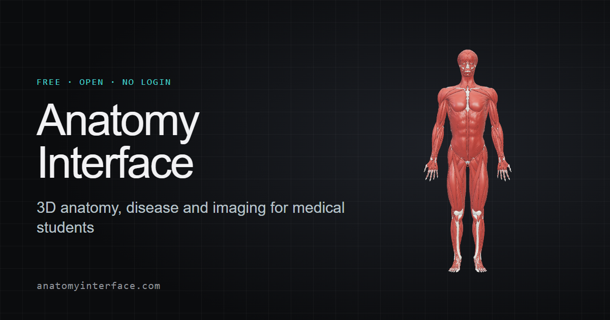
Last built 2026-11-13.